

Translate this page into:
A Case of Chylothorax in a Hemodialysis Patient with Left Innominate Venous Stenosis
This is an open access article distributed under the terms of the Creative Commons Attribution-NonCommercial-ShareAlike 3.0 License, which allows others to remix, tweak, and build upon the work non-commercially, as long as the author is credited and the new creations are licensed under the identical terms.
This article was originally published by Medknow Publications & Media Pvt Ltd and was migrated to Scientific Scholar after the change of Publisher.
Abstract
Chylothorax is defined as accumulation of chyle-containing lymphatic fluid within the pleural space. Chylothorax is very rarely seen in hemodialysis patients. We report a case of a patient on hemodialysis who developed chylothorax secondary to left innominate vein stenosis, with other features of venous hypertension such as arm edema successfully treated with angioplasty and pigtail drainage.
Keywords
Chylothorax
dialysis
innominate vein stenosis

Introduction
Chylothorax is defined as accumulation of chyle-containing lymphatic fluid within the pleural space. The causes of chylothorax are various and are usually attributable to one of the categories: malignancy, trauma (including surgery), miscellaneous disorders, and idiopathic. Occurrence of chylothorax in hemodialysis is very uncommon, and it may have resulted from iatrogenic vasculature trauma conducive to venous thrombosis and stenosis when hemodialysis catheter requires frequent change or long-term indwelling. Local thrombosis and stenosis may increase the venous hydrostatic pressure and hinder the drainage of the thoracic lymph duct into the venous system, hence chylous lymphatic fluid leak into the pleural space.
Case Report
A 45-year-old male patient with end-stage renal disease secondary to obstructive nephropathy was admitted with complaint of the left neck, left half face, and left upper limb swelling of 2 months duration. He denied recent trauma, fever, or chills. Medications at the time of presentation were amlodipine, pantoprazole, carvedilol, calcium carbonate, and calcitriol. The patient had begun hemodialysis for 1½ years before the current admission through a left internal jugular catheter. The patient denied the history of fever, chest pain, night sweats, or significant weight loss, suggestive of pulmonary or extrapulmonary tuberculosis. He had primary left radiocephalic arteriovenous (AV) fistula failure; hence, a left brachiocephalic AV fistula was created and following which he developed severe venous hypertension of the left upper limb.
On physical examination, patient was afebrile with blood pressure of 140/90 mmHg. He had diffuse edema over the left half of his face, neck, chest, and left upper limb. AV fistula was not infected. There was no evidence of thyroid or any lymph node swelling. His remaining physical examination was unremarkable; no jugular venous distension, murmurs or rubs on cardiac examination, clear lungs were reported.
Laboratory evaluation showed hemoglobin of 10.4 g/dl, total count of 8400 cells/mm3 with normal liver function tests, and bleeding time and clotting time were within normal limits. T3, T4, and thyroid-stimulating hormone were within normal limits. Chest X-ray showed increased density and swelling of soft tissue overlying the left neck, chest and arm with massive left-sided pleural effusion. Doppler of the neck vessels showed decreased flow in the left subclavian vein. Echocardiography showed normal biventricular functions with ejection fraction of 62%. Computerized tomography (CT) of the thorax showed left massive pleural effusion but no evidence of any malignancy or lymphadenopathy; after obtaining the informed consent, the patient underwent conventional angiogram which showed left innominate vein total occlusion [Figure 1]. Angioplasty of the left innominate vein was done [Figure 2], following which his facial and upper limb swelling drastically reduced [Figure 3].

- Angiogram showing left innominate vein stenosis

- Angiogram showing postangioplasty balloon dilatation
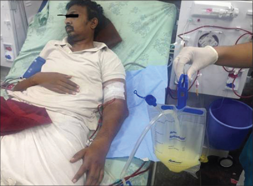
- Patient with severe venous edema of the left upper limb and chylous pleural effusion
A pig-tail was inserted into left pleural space and 1 L of milky fluid was drained. Laboratory analysis of the pleural fluid showed triglycerides of 990 mg/dl and lymphocyte predominance (90%) on cell count differential consistent with chyle. All the other causes of pleural effusion were ruled out with echocardiography and CT of the thorax. Pleural fluid cultures were negative for bacteria, fungus, and acid-fast bacilli. The patient continued to drain 500–600 ml/day of chyle, and by 1 week, the drain reduced to <50 ml and pigtail was removed. A repeat chest X-ray showed complete resolution of left-sided pleural effusion.
Discussion
We describe a case of end-stage renal disease patient on hemodialysis who presented with pleural effusion with severe left upper limb venous hypertension.[1] Chylothorax can be left-sided (33.3%) when due to damage above the fifth thoracic vertebra, right-sided (50%) when due to thoracic duct damage below this level, or bilateral (16.7%).[2] Other causes include malignancy, cardiac failure, sarcoidosis, goiter, as well as thoracic and upper gastrointestinal surgery.
Our patient had severe left innominate vein stenosis which was the probable etiology of chylous pleural effusion, which is in turn related to previous dialysis catheter insertion. The left innominate vein stenosis will increase the pressure in the thoracic duct and cause subsequent chylous leak into the pleural cavity causing chylothorax. There are very few case reports of chylous pleural effusion with superior vena caval (SVC) obstruction.[1]
Less than 50% of chylothoraces have the classic milky appearance,[3] so diagnosis depends on pleural fluid analysis showing triglyceride concentrations >110 mg/dl (or the detection of chylomicrons). Lymphography can be considered for localization of thoracic duct obstruction.[4] Treatment of the underlying cause is crucial, often resulting in spontaneous cessation of chyle leakage. With a leak, thoracic duct ligation may be preferred, especially for a traumatic cause. Octreotide has been used with varying success. Ceasing fat intake will reduce chyle formation, requiring parenteral supplement of medium chain fatty acids. Thoracocentesis is beneficial for symptom relief but should be done cautiously to avoid immunosuppression from loss of chyle. Infections rarely occur in the pleural effusion itself since chyle is bacteriostatic. The chylous pleural effusion was completely treated by combined angioplasty and pleural drainage.
Conclusion
Chylothorax must be considered as one of the causes of pleural effusion in hemodialysis patients with severe venous hypertension of the upper limbs. Although rare, a successful treatment of chyle pleural effusion can be achieved with the treatment of venous stenosis in the form of angioplasty and pleural drainage.[5]
Declaration of patient consent
The authors certify that they have obtained all appropriate patient consent forms. In the form the patient(s) has/have given his/her/their consent for his/her/their images and other clinical information to be reported in the journal. The patients understand that their names and initials will not be published and due efforts will be made to conceal their identity, but anonymity cannot be guaranteed.
Financial support and sponsorship
Nil.
Conflicts of interest
There are no conflicts of interest.
References
- Chylothorax: Aetiology, diagnosis and therapeutic options. Respir Med. 2010;104:1-8.
- [Google Scholar]
- Chylothorax and chylopericardial tamponade in a hemodialysis patient with catheter-induced superior vena cava stenosis. Semin Dial. 2009;22:576-9.
- [Google Scholar]
- The lipoprotein profile of chylous and nonchylous pleural effusions. Mayo Clin Proc. 1980;55:700-4.
- [Google Scholar]
- Chylous pericardial tamponade in a haemodialysis patient with catheter-associated thrombosis of internal jugular and subclavian veins. Nephrol Dial Transplant. 2006;21:2650-3.
- [Google Scholar]


