

Translate this page into:
Hemodialysis-related Portal-systemic Encephalopathy: A Rare Cause of Recurrent Encephalopathy among Patients on Maintenance Hemodialysis
-
Received: ,
Accepted: ,
This article was originally published by Wolters Kluwer - Medknow and was migrated to Scientific Scholar after the change of Publisher.
Abstract
Portal-systemic venous shunts can rarely develop without any intrinsic liver diseases. However, the cause of shunt formation in these cases are not very clear. Literature suggests that hemodialysis can precipitate symptoms in patients with asymptomatic portal-systemic venous shunts (PSVS). Rare presentations of recurrent encephalopathy due to PSVS in the absence of liver dysfunction has been described in patients undergoing hemodialysis. We report a rare case of recurrent Hemodialysis Related Porto-Systemic Encephalopathy (HRPSE) in a 50-year old male during maintenance hemodialysis secondary to a PSVS between the portal vein and left renal vein. Shunt embolism by an 18 mm Amplatzer vascular plug (AVR II) was done and follow up CT showed complete occlusion of collaterals. Post-procedure, he is undergoing thrice-weekly Hemodialysis of 4 hours duration till date with no further incidence of encephalopathy. Our report indicates that recurrent encephalopathy can occur in dialysis patients due to symptomatic PSVS and HRPSE should be considered even in non-cirrhotic cases for early detection and effective management.
Keywords
Hemodialysis
portal-systemic venous shunts
shunt embolism

Introduction
Portal-systemic venous shunts (PSVS) are usually formed by portal hypertension associated with cirrhosis, which often results in recurrent hepatic encephalopathy accompanied by hyperammonaemia.[1] However, in a minority of cases PSVS can develop without intrinsic liver disease, and the cause of shunt formation in these patients is not clear.[2] Among dialysis population, among various causes of encephalopathy, PSVS are listed as a rare cause, and literature suggests hemodialysis as a risk factor in precipitating symptoms in patients with asymptomatic PSVS.[3] Rare presentations of recurrent encephalopathy due to PSVS in the absence of liver dysfunction has been described in patients undergoing hemodialysis-Hemodialysis Related Porto-Systemic Encephalopathy (HRPSE).[4] We report a rare case of recurrent HRPSE in a 50-year old male during maintenance hemodialysis secondary to a PSVS between the portal vein and left renal vein and he improved remarkably with shunt embolisation.
Case History
A 50-year-old gentleman with history of DM with microvascular complications, was initiated on hemodialysis through right IJV cuffed catheter following irreversible acute on chronic kidney disease due to superadded “Infection-related GN” on underlying advanced diabetic nephropathy. He had low C3 levels and kidney biopsy had shown diffuse proliferative glomerulonephritis with background diabetic nodular glomerulosclerosis, 5 of 13 glomeruli showed cellular crescents. The renal function progressively deteriorated despite trial of steroids. His past history was significant for an episode of acute pancreatitis, acute coronary syndrome on medical management and regular alcohol consumption.
Two months into HD he developed acute onset altered mental status. On examination, he had gait instability and flaps. His serum Ammonia level was elevated (67 mmol/L) despite acceptable urea reduction rates on hemodialysis. Ultrasound abdomen showed fatty liver and upper GI endoscopy showed no varices. Liver stiffness measurement done using Echosens FibroScan 402 showed a medium stiffness value of 7.5 kPa which ruled out significant fibrosis. Brain imaging was normal. He was treated with intensive hemodialysis and hepatic coma measures following which he recovered. His access was changed to left forearm AVF. Five months later he presented with second episode of altered mental status in the form of acute onset agitation which rapidly progressed to stupor within 4 hours. Repeat brain imaging was normal, but serum ammonia was elevated (104 mmol/L). He was treated with high-dose thiamine, hepatic coma measures and intensification of hemodialysis. His sensorium gradually improved over a day. He needed two more admissions for similar complaints 6 and 11 months after initiation of HD with ammonia levels of 246 mmol/L and 291 mmol/L respectively and improved with conservative measures and intensification of dialysis as in prior episodes. Biochemical parameters 2 months, 5 months, 6 months, and 11 months after hemodialysis initiation are summarized in Table 1. In the absence of advanced cirrhosis and lack of clinically significant portal hypertension a contrast CT of abdomen was done to look for any abnormal portosystemic communication. A tortuous and prominent collateral vein from the portal vein was seen draining to the left renal vein [Figures 1 and 2]. Shunt embolism by an 18 mm Amplatzer vascular plug (AVR II) was done on October-2019 [Figure 3]. Follow up CT showed complete occlusion of collaterals [Figure 4]. He was put on oral anticoagulation in view of thrombosis of common iliac and right external iliac veins after procedure. His serum ammonia level three days after the procedure was 37 μmol/L. Post-procedure, he is undergoing thrice-weekly Hemodialysis of 4 hours duration till date with no further incidence of encephalopathy.
| Parameters | 2 months after HD initiation | 5 months after HD initiation | 6 months after HD initiation | 11 months after HD initiation |
|---|---|---|---|---|
| Urea (mg/dl) | 67 | 91 | 75 | 101 |
| Ammonia (µmol/L) | 67 | 104 | 246 | 291 |
| Sodium (meq/L) | 133 | 137 | 138 | 133 |
| Ionised Ca (mg/dl) | 1.00 | 0.87 | 1.05 | 1.12 |
| HIV/HCV/HBsAg | Non-reactive | Non-reactive | Non-reactive | Non-reactive |

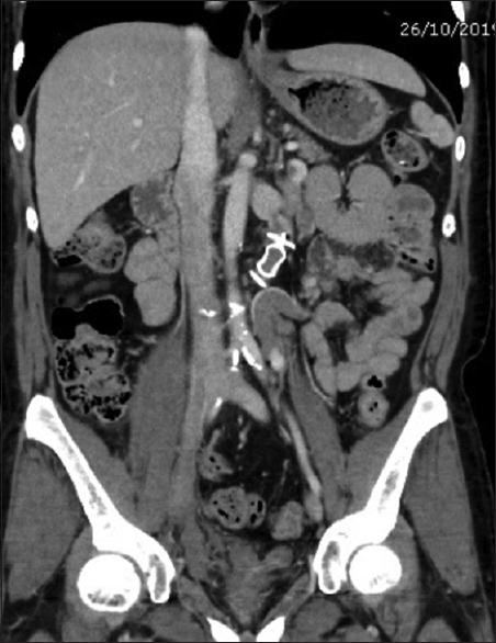
- Contrast CT abdomen before the procedure

- 3D reconstructed image of shunt (Red) between portal vein and left renal vein
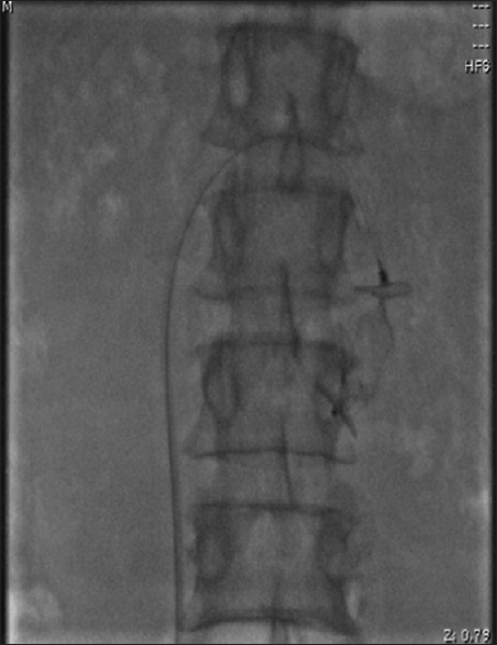
- Fluoroscopic image just after deploying Amplatzer vascular plug
- CECT abdomen after embolisation of the portal-systemic shunt using Amplatzer vascular plug
Discussion
We report a patient with end-stage renal disease from advanced diabetic nephropathy who developed hepatic encephalopathy because of the presence of a PSVS between the portal vein and left renal vein. The patient experienced no episodes of encephalopathy after shunt embolization.
Recurrent encephalopathy caused by spontaneous PSVS in patients undergoing hemodialysis was reported rarely in the past. Shimono et al. described the case of a 57-year-old male with diabetic nephropathy managed with maintenance hemodialysis who developed recurrent encephalopathy due to a PSVS from the superior mesenteric vein via the left gastric vein to the left renal vein. The patient symptomatically improved after undergoing a shunt ligation.[5] However, this patient had a past history of a partial gastrectomy which is an independent risk factor for a PSVS.[6] Another case report from Japan described the case of a 68-year-old woman who developed encephalopathy after her ninth session of hemodialysis and investigations (Color Doppler ultrasonography and magnetic resonance angiography) revealed a PSVS between the left gastric vein and left renal vein. She improved with surgical ligation of the shunt.[3] Another case was reported by Kondo et al. where a 75-year-old woman developed encephalopathy five years after the initiation of hemodialysis secondary to a large PSVS between the left splenic and left renal veins and she was treated with catheter therapy to occlude the abnormal shunt vessel.[4]
Hemodialysis-related portal-systemic encephalopathy (HRPSE) in patients without chronic liver disease is rare, and the pathogenesis of this disease remains unclear. A recent study analyzed 13 reports of HRPSE and found that most patients were Asian women in their sixties.[4] There are multiple explanations for the PSVS; the persistence of portal–hepatic venous systems during embryonic development, spontaneous development, or a sequelae of external abdominal injury or surgery.[57] It usually go unnoticed due to absence of symptoms. However, various factors such as aging, constipation, high protein diet, or hemodynamic treatment of hemodialysis can precipitate symptoms in patients with PSVS.[38] Ubara et al. in their report hypothesized that rapid removal of a large fluid volume in anuric patient during hemodialysis transiently reduced total body fluid volume leading to reduced venous pressure, thereby increasing portal venous blood flow into the inferior vena cava via PSVS, resulting in ammonia-rich blood reaching the systemic circulation, producing encephalopathy.[3]
The treatment of HRPSE is the occlusion of the PSVS. Surgical correction of PSVS was reported since 1982.[5] Recent advances in interventional radiology had shown effective treatment of HRPSE such as trans-portal obliteration of shunts, balloon-occluded retrograde trans-venous obliteration, and shunt-preserving disconnection of portal and systemic circulation, to occlude the abnormal vessels.[1] However, embolization therapy using micro coils could not be performed in cases with large shunt, where surgical ligation of the shunt is advocated.[3]
In conclusion, we reported a patient with recurrent HRPSE secondary to a PSVS between the portal vein and left renal vein who improved markedly from embolization of the shunt. Our report indicates that recurrent encephalopathy can occur in dialysis patients due to symptomatic PSVS and HRPSE should be considered even in non-cirrhotic cases for early detection and effective management.
Declaration of patient consent
Written informed consent was obtained from the patient for the publication of this case report. The patients understand that their names and initials will not be published and due efforts will be made to conceal their identity, but anonymity cannot be guaranteed.
Financial support and sponsorship
Nil.
Conflicts of interest
There are no conflicts of interest.
References
- Portal-systemic encephalopathy in non-cirrhotic patients: Classification of clinical types, diagnosis and treatment. J Gastroenterol Hepatol. 2000;15:969-79.
- [Google Scholar]
- Spontaneous intrahepatic portal-systemic venous shunt in the adult: Case report and review of the literature. Dig Dis Sci. 2004;49:1201-6.
- [Google Scholar]
- Hemodialysis-related portal-systemic encephalopathy. Am J Kidney Dis. 2004;44:e38-42.
- [Google Scholar]
- Recurring encephalopathy abolished by gastrorenal shunt ligation in a diabetic hemodialysis patient. Am J Gastroenterol. 1998;93:270-2.
- [Google Scholar]
- Case report: An unusual case of portal systemic encephalopathy. Am J Dig Dis. 1975;20:176-81.
- [Google Scholar]
- Portal-systemic encephalopathy due to a congenital portocaval shunt. Am J Radiol. 1982;139:1013-5.
- [Google Scholar]
- Portal systemic encephalopathy in a non-cirrhotic patient. J Neurol Neurosurg Psychiatry. 2008;79:96.
- [Google Scholar]


