Translate this page into:
Color Doppler findings of post-biopsy arteriovenous fistula in renal transplant
Dr. Aijaz Hakeem, Department of Radio-Diagnosis, SK Institute of Medical Sciences, Jammu and Kashmir - 190 011, India. E-mail: aijazhakeem@yahoo.com
This is an open-access article distributed under the terms of the Creative Commons Attribution License, which permits unrestricted use, distribution, and reproduction in any medium, provided the original work is properly cited.
This article was originally published by Medknow Publications and was migrated to Scientific Scholar after the change of Publisher.
Abstract
Post biopsy arterio-venous fistula in renal transplant range in incidence from 15-16%. Spontaneous resolution of 75% A-V fistulas is seen within four weeks. We report a patient with post biopsy arterio-venous fistula who had developed unexplained hypertension with no definite feature of rejection on biopsy. Doppler application revealed an arterio-venous fistula which showed spontaneous resolution in six weeks.
Keywords
Arteriovenous fistula
color Doppler
post-biopsy
Introduction
In renal transplants post biopsy fistulas are well known. Most resolve spontaneously only few which are large need intervention. We report a case in whom we had done renal biopsy and who developed post biopsy AV fistula which we documented on color Doppler. He was followed for 6 weeks after which we noticed a spontaneous resolution. Most of the post biopsy AV fistula need follow up and are likely to resolve after some period.
Case Report
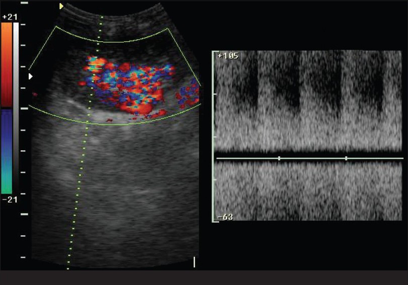
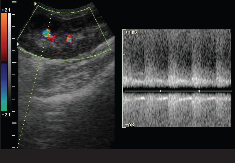
We evaluated a 46-year-old male, who had received live donor renal transplant one year ago, for a recent onset of hypertension. We performed three renal biopsies over a period of three months with no evidence of chronic transplant rejection. The patient was referred to the Radiology department for ultrasonography (USG) Doppler of the transplant. We performed a color Doppler study of the transplant kidney, which revealed an arteriovenous (AV) fistula in the lower pole involving lobar vessels with markedly increased peak systolic velocity (PSV; >170 cm/s), end diastolic velocity (EDV; >70 cm/s) and a reduced resistive index (RI = 0.45). Arterialization of the venous waveform was evident along with perivascular random color assignment [Figs. 1 and 2]. The renal artery and vein at the hilum appeared normal. A follow-up after 6 weeks showed spontaneous resolution of the fistula.

- Showing turbulent flow on pulse wave doppler study

- Arterializations of the venous waveform along with peri-vascular random color assignment on pulse wave Doppler flow
Discussion
The incidence of post-biopsy renal transplant AV fistulas range from 15% to 16%.12 Only a small percentage of these fistulas are sufficiently large in size to warrant surgical intervention for closure.34 Few cases are associated with pseudoaneurysms, which if small in size, resolve simultaneously with the fistula.2 The AV fistulas are characterized by a region of high velocity shifts with random color assignment due to vibrating interfaces in the perivascular tissue5 and arterialization of venous flow, the latter distinguishing it from focal renal artery stenosis.1 Most of the complications occur after renal biopsy, including AV fistulas. The AV fistulas are occluded by transcatheter embolotherapy wherein a steel coil is placed into the fistula from the renal vein approach. This procedure permits nonsurgical closure of the AV shunt without significantly altering the renal function.6 Pseudoaneurysms are treated conservatively.7 A spontaneous resolution of 75% in AV fistulas was noted within four weeks. All pseudoaneurysms located close to the AV fistulas are also spontaneously closed. In most cases, post-biopsy AV fistulas are clinically occult and resolve spontaneously in 1–2 years.8
In most cases, color and duplex Doppler USG easily demonstrate AV fistulas besides other obvious vascular and nonvascular complications and requires invasive procedures such as renal angiography.
Source of Support: Nil
Conflict of Interest: None declared.
References
- Postbiopsy renal transplant arteriovenous fistulas: Color Doppler US characteristics. Radiology. 1989;171:253-7.
- [Google Scholar]
- Color coded duplex sonographic study of arteriovenous fistulas and pseudo aneurysms complicating percutaneous renal allograft biopsy. Clin Nephrol. 2002;58:398-404.
- [Google Scholar]
- Renal allograft arteriovenous fistula and large pseudo aneurysm. Clin Transplant. 2003;17:9-12.
- [Google Scholar]
- Color flow Doppler imaging of carotid artery abnormalities. AJR Am J Roentgenol. 1988;150:419-25.
- [Google Scholar]
- Arteriovenous fistula after biopsy of renal transplant kidney: Diagnosis and treatment. Pediatr Nephrol. 1992;6:562-4.
- [Google Scholar]
- Intrarenal arteriovenous fistulas following needle biopsy of the kidney. J Paediatr. 1971;78:266-72.
- [Google Scholar]


