Translate this page into:
Peculiar Acral Melanosis after Cyclophosphamide Therapy in a Case of Primary Membranous Nephropathy: A Rare Presentation
Address for correspondence: Dr. Karthikeyan Balasubramanian, Department of Nephrology, Saveetha Medical College Hospital, Thandalam, Chennai - 602 105, Tamil Nadu, India. E-mail: karthiprakash1986@gmail.com
-
Received: ,
Accepted: ,
This is an open access journal, and articles are distributed under the terms of the Creative Commons Attribution-NonCommercial-ShareAlike 4.0 License, which allows others to remix, tweak, and build upon the work non-commercially, as long as appropriate credit is given and the new creations are licensed under the identical terms.
This article was originally published by Wolters Kluwer - Medknow and was migrated to Scientific Scholar after the change of Publisher.
Dear Sir,
Alkylating agent like cyclophosphamide has been utilized in the management of membranous nephropathy as a part of the modified Ponticelli regimen. Although well tolerated by the majority of the patients, various adverse events have been reported commonly (>10%) like GI disturbances, haematological side effects like cytopenias, alopecia, and amenorrhoea. Cutaneous manifestations of cyclophosphamide therapy are rarely reported. Hyperpigmentation, both diffuse and acral, is a common adverse effect of various anticancer drugs.[12] Cyclophosphamide-induced hyperpigmentation usually occurs on the photo-exposed skin by a photomediated pathogenetic mechanism.[34]
A 47-years-old non-diabetic, normotensive lady presented in our Nephrology outpatient department with complaints of pedal oedema and facial puffiness for 6 months, which was insidious in onset, gradually progressive. Her examination showed blood pressure of 130/80 mmHg with pitting type of pedal oedema. Her urine examination showed 3+ albumin on dipstick with bland urinary sediments. At the onset of symptoms 6 months back, her investigations revealed urine showing +3 albumin and 24-hour urinary protein of 4.5 gm/day with dyslipidemia and severe hypoalbuminemia. She underwent renal biopsy and diagnosed to have Primary Membranous Nephropathy, which showed IF IgG (+3), C3 (+1); granular positivity over capillary loops and tissue staining for PLA2R (+) and was diagnosed as primary Membranous Nephropathy. She was treated with RAAS blockade therapy, diuretics and statins for 6 months. As her symptoms did not improve she was referred to our hospital. Her investigations revealed urine spot Protein creatinine ratio (6.12), S. Albumin (2.3 g/dl), and S. Creatinine (0.9 mg/dl). After obtaining consent, she was started on modified Ponticelli regimen. The patient was given 1 gm of methylprednisolone for three doses followed by oral prednisolone 0.5 mg/kg/day for 27 days. Next month she was started on 2 mg/kg/day of oral cyclophosphamide.
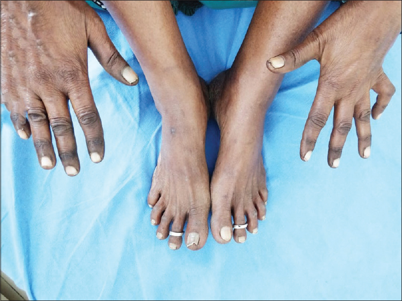
During the follow-up, her renal functions were stable, proteinuria improved and she had attained partial remission (Urine spot PCR 2.73 & S. Albumin 2.6g/dl) at the end of the second cycle. During the treatment with cyclophosphamide, she developed dark brown hyperpigmentation involving bilateral ventral and dorsal aspects of hand with more involvement over the index finger and thumb. The pigmentation was more in the tip of the fingers, knuckles. Similar hyperpigmentation is also seen in bilateral dorsum of feet and toes. Nail involvement was not present [Figures 1 and 2]. She was otherwise asymptomatic and afebrile. These complaints started after 3 weeks of cyclophosphamide therapy. There was no history of redness, pain, and swelling over this area. Upon examination temperature was normal in all four limbs, not associated with any tenderness, bilaterally her peripheral pulses were equal in both upper and lower limbs, and no evidence of distal neurovascular deficit or Raunaud's phenomenon was observed. AV Doppler of all four limbs was done, which showed normal flow. Investigations showed leucocytopenia (TC-2600/mm3) with normocytic normochromic RBCs. Considering the possibility of autoimmune association with Membranous nephropathy; serum ANA profile (immunoblot) was done which was negative. ESR and CRP were normal. Her vitamin B12 levels, thyroid profile were normal. There are no features of Addison disease like mucosal pigmentation, hypotension, and electrolyte abnormalities. After excluding these conditions, she was diagnosed as drug-induced acral melanosis secondary to cyclophosphamide therapy. We reduced the dose of oral cyclophosphamide to 50 mg/day. Her pigmentation decreased during follow-up over 1 month. When she was started on cyclophosphamide in the fourth month, she again had mild increase in pigmentation associated with low WBC count that was managed conservatively. Thus a provisional diagnosis of cyclophosphamide-induced acral melanosis in Primary Membranous nephropathy on modified Ponticelli regimen was considered.

- Diffuse acral hyperpigmentation involving palmar surface of both hands with pigmentation more at tip of fingers

- Acral pigmentation involving dorsum of hands, knuckles, and feet
The causes of acral pigmentation varies from genetic to acquired, benign to malignant, autoimmune to infectious, drug induced, nutritional deficiencies, post inflammatory, and even exogenous reasons.[1] The pigmentation can occur in isolation or can be associated with various systemic features. An earlier age of onset of pigmentation, a positive family history, and a reticulate or mottled pattern usually point to a genetic cause while diffuse pattern of pigmentation as seen is usually seen in racial, drug induced, endocrine diseases, and nutritional deficiencies.[5] The drugs that can cause hyperpigmentation, which are used frequently in patients with kidney disease are hydroxychloroquine, ketoconazole, and tetracycline antibiotics.[1] Angiotensin receptor blockers, steroids, calcium supplements used in nephrotic syndrome patients do not usually cause acral hyperpigmentation.
Our patient had diffuse pattern of acquired acral hyperpigmentation following cyclophosphamide therapy with cytopenia in the background of Membranous nephropathy; a detailed evaluation for autoimmune association or Raynaud's phenomenon was performed. However, there was no evidence of inflammatory/autoimmune features/nutritional deficiencies/gastrointestinal involvement, which could be attributed to acral hyperpigmentation. Therefore, a provisional diagnosis of acral melanosis due to dose-dependent effects of cyclophosphamide therapy used as a part of the modified Ponticelli regimen was considered, which improved on dose reduction or stoppage of the drug. Hence, the final diagnosis of Cyclophosphamide-induced acral melanosis was established.
The caveats in the diagnosis of acral melanosis due to cyclophosphamide therapy is its rare occurrence and under reporting in real-world setting. There are only 10%-20% cases of drug-induced (acquired) acral pigmentation reported worldwide; however, a case report with the use of cyclophosphamide in modified Ponticelli regimen is yet to be reported.
Declaration of patient consent
The authors certify that they have obtained all appropriate patient consent forms. In the form the patient has given her consent for her images and other clinical information to be reported in the journal. The patient understands that her name and initials will not be published and due efforts will be made to conceal her identity, but anonymity cannot be guaranteed.
Financial support and sponsorship
Nil.
Conflicts of interest
There are no conflicts of interest.
References
- Drug-induced skin pigmentation.Epidemiology, diagnosis and treatment. Am J Clin Dermatol. 2001;2:253-62.
- [Google Scholar]


