Translate this page into:
Tunneled Hemodialysis Catheter-associated Right Atrial Thrombus Presenting with Septic Pulmonary Embolism
Address for correspondence: Dr. S. V. Vyahalkar, Department of Nephrology, Grant Medical College and Sir JJ Group of Hospitals, Mumbai, Maharashtra, India. E-mail: drsameer2010@gmail.com
This is an open access journal, and articles are distributed under the terms of the Creative Commons Attribution-NonCommercial-ShareAlike 4.0 License, which allows others to remix, tweak, and build upon the work non-commercially, as long as appropriate credit is given and the new creations are licensed under the identical terms.
This article was originally published by Medknow Publications & Media Pvt Ltd and was migrated to Scientific Scholar after the change of Publisher.
Abstract
Tunneled hemodialysis (HD) catheter-associated right atrial thrombus (CRAT) is an uncommon complication with significant morbidity. We report the case of a patient undergoing HD through tunneled venous catheter who presented with catheter dysfunction and sepsis and was diagnosed to have CRAT with septic embolism. CRAT formation has a significant association with catheter-related infection. The need for early diagnosis and various treatment options for this entity are highlighted.
Keywords
Catheter-associated right atrial thrombus
pulmonary embolism
sepsis
tunneled hemodialysis catheter
Introduction
Central venous catheter-associated right atrial thrombus (CRAT) is a well-documented complication in patients receiving chemotherapy and parenteral nutrition; however, data regarding CRAT in hemodialysis patients with tunneled venous catheters (TVCs) are limited. CRAT in hemodialysis (HD) patients is a recognized complication when TVC is left in place for a prolonged duration. It generally presents with catheter dysfunction and can lead to potentially life-threatening consequences like pulmonary thromboembolism, septic emboli, endocarditis, septic or cardiogenic shock and cardiac arrest if not diagnosed early. The management of CRAT in TVCs is highly individualized due to the associated comorbidities.
Case Report
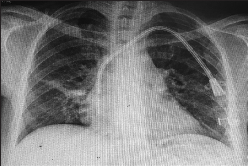
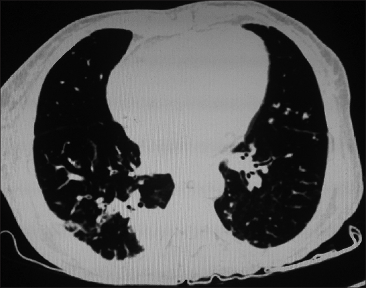
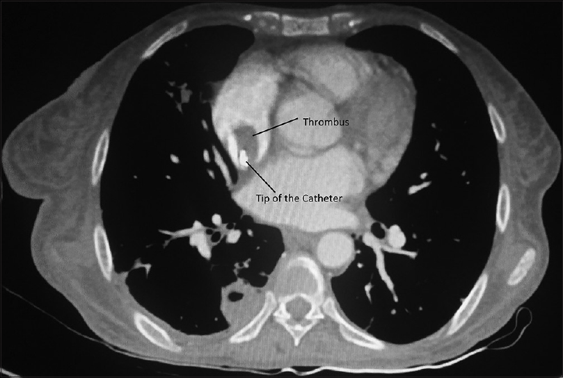
A 51-year-old female patient with end-stage renal disease, undergoing HD for 1 year presented with complaints of malaise, fever, and dry cough for 1 month. She had a history of two failed arterio-venous fistula (AVF) surgeries, and due to temporary HD catheter-related thrombosis of the right internal jugular vein (IJV), TVC was inserted in left IJV 10 months ago. Poor blood flow from catheter and intra-dialysis fever with rigors was noted and she was being treated for suspected catheter-related bloodstream infection (CRBSI) at the dialysis unit for the past 2 weeks. Four months before present admission, she was hospitalized for suspected CRBSI and cuff extrusion and TVC was removed. Blood or catheter culture study was not done at that time; however, she had been treated with intravenous antibiotics and TVC (23 cm Hemosplit® BARD Inc.) was re-inserted in left IJV. On examination, she was febrile, had tachycardia, hypotension (systolic BP 90 mmHg) and had signs of protein calorie malnutrition. Investigations revealed Hb 6.6 g/dl, white blood cell 15,900/cmm, platelets 107,000/cmm, serum creatinine 12.3 mg/dl, blood urea 167 mg/dl, and serum albumin 2.6 g/dl. Blood culture was negative (while on antibiotics). X-ray chest revealed diffuse bilateral infiltrates with the tip of TVC in the right atrium (RA) [Figure 1]. Sputum examination, including staining for acid-fast bacilli and cartridge-based nucleic acid amplification test (GeneXpert® MTB/RIF) for Mycobacterium tuberculosis was negative. Sputum staining for fungi, Gram staining and bacterial culture were also negative. Contrast enhanced computed tomography of thorax revealed partial contrast filling defect in segmental branches of both lower lobes and diffuse centrilobular nodules bilaterally, few of which coalesced to form areas of consolidation with cavities [Figure 2] and no evidence of lymphadenopathy. Also noted was a large hypodense filling defect around the TVC in RA [Figure 3]. Therefore, transthoracic echocardiography (TTE) was done, which revealed a mobile thrombus measuring 4 cm × 1.1 cm around the tip of TVC extending to the tricuspid valve causing ball valve effect, without significant tricuspid regurgitation or pulmonary hypertension. These features suggested CRAT and septic pulmonary embolism. She was treated with antibiotics (meropenem and vancomycin) and unfractionated heparin. Noncuffed catheter was secured in the femoral vein for HD and TVC removal was planned after attaining therapeutic anticoagulation; however, the patient took discharge against medical advice and lost to follow-up.

- X - ray chest showing tip of TVC in right atrium and bilateral diffuse infiltrates

- Computed tomography thorax reveals bilateral diffuse centrilobular nodules with cavitation in basal segments

- Contrast-enhanced computed tomography thorax shows filling defect adjacent to the catheter tip in right atrium
Discussion
Factors responsible for the development of CRAT include activation of coagulation cascade following catheter-induced endothelial injury, blood flow changes associated with localization of catheter tip (in the RA rather than superior vena cava)[1] and the hypercoagulable state in chronic kidney disease[2] besides conventional risk factors for thrombogenesis. The incidence of CRAT in dialysis patients with TVCs is not precisely known because most of the data come from small case series. A single center 3 years audit of 100 TVCs with a cumulative follow-up of 492 patient months from India[3] did not report the occurrence of CRAT. In a retrospective case series, frequency of CRAT was estimated to be 5.4%;[4] whereas, a higher frequency of 18% was found in a study[5] that used TTE to look for the presence of CRAT in dialysis patients with TVCs, implying that asymptomatic CRAT is not uncommon.
There is a significant association between CRAT and CRBSI, but the precise etiopathological relationship is unclear.[46] Even though a catheter is removed following CRBSI, catheter-related infective nidus may remain and act as focus for development of CRAT once new catheter is inserted.[7] This may also explain our patient's course. Therefore, active surveillance for CRAT is necessary in patients who require repeated TVC insertion. Urgent echocardiography should be considered to exclude an infected CRAT in catheter-dependent HD patients presenting with sepsis,[8] and is the most commonly used imaging modality for diagnosis of CRAT[9] with the advantages of low cost, noninvasiveness, and wide availability.
Optimal management of CRAT remains controversial for lack of adequate data. Catheter removal after attaining therapeutic anticoagulation is the treatment in most of the cases with a diagnosis of CRAT.[9] Early removal of TVC is considered in the presence of bacteremia[4] and nonadherent small thrombi. If large adherent thrombi are present, the TVC is left in place for some duration till regression of thrombus is achieved with therapeutic anticoagulation before its removal. Patients are usually treated with anticoagulation using INR target of 2–3 for 6 months; the duration of anticoagulation may vary depending on the extent of thrombosis and need for continued use of the catheter.[9] However, anticoagulation carries a higher risk of bleeding complications in dialysis population, and there are no randomized controlled trials of anticoagulation in dialysis population to recommend its use.[10] Stavroulopoulos et al. formulated a treatment plan for CRAT associated with TVCs based on meta-analysis of case reports, and recommend removal of TVC and anticoagulation for patients with a thrombus size <6 cm; whereas surgical thrombectomy is recommended in those with a thrombus >6 cm, contraindication to anticoagulation or cardiac abnormalities like endocarditis. Catheter-directed or systemic thrombolysis, although seldom successful in CRAT,[9] is recommended in managing hemodynamically unstable right heart pulmonary thromboembolism.[11] This case presented therapeutic challenges like a high surgical risk for thrombectomy and technical limitations of percutaneous interventions, which restricted the therapeutic choices, risk of early removal of TVC (i.e., pulmonary thromboembolism) and at the same time, risk of therapeutic anticoagulation. It is important to maintain a high index of suspicion for CRAT and thrombo-embolic complications in dialysis patients with TVCs who present with catheter dysfunction or catheter-related infection and echocardiography should be considered for early diagnosis to reduce morbidity and mortality. The case highlights the importance of limiting the duration of catheter use and timely placement of permanent arteriovenous access.
Financial support and sponsorship
Nil.
Conflicts of interest
There are no conflicts of interest.
References
- Atrial thrombus and central venous dialysis catheters. Am J Kidney Dis. 2001;38:631-9.
- [Google Scholar]
- Tunneled central venous catheters: Experience from a single center. Indian J Nephrol. 2011;21:107-11.
- [Google Scholar]
- Right atrial thrombi complicating use of central venous catheters in hemodialysis. J Vasc Access. 2005;6:18-24.
- [Google Scholar]
- The relationship between the thrombotic and infectious complications of central venous catheters. JAMA. 1994;271:1014-6.
- [Google Scholar]
- Multidetector row CT diagnosis of an infected right atrial thrombus following repeated dialysis catheter placement. Br J Radiol. 2009;82:e240-2.
- [Google Scholar]
- Right atrial mass related to indwelling central venous catheters in patients undergoing dialysis. Eur J Echocardiogr. 2000;1:222-3.
- [Google Scholar]
- Right atrial thrombi complicating haemodialysis catheters. A meta-analysis of reported cases and a proposal of a management algorithm. Nephrol Dial Transplant. 2012;27:2936-44.
- [Google Scholar]
- Sailing between Scylla and Charybdis: Oral long-term anticoagulation in dialysis patients. Nephrol Dial Transplant. 2013;28:534-41.
- [Google Scholar]
- Right heart thrombi in pulmonary embolism: Results from the International Cooperative Pulmonary Embolism Registry. J Am Coll Cardiol. 2003;41:2245-51.
- [Google Scholar]


