

Translate this page into:
Acute Kidney Injury Secondary to Leukemic Infiltration of the Kidneys in M3 Acute Myeloid Leukemia

-
Received: ,
Accepted: ,
How to cite this article: Aggarwal J, Pathak NM, Rathore V, Badge RP, Sharma A. Acute Kidney Injury Secondary to Leukemic Infiltration of the Kidneys in M3 Acute Myeloid Leukemia. Indian J Nephrol. doi: 10.25259/IJN_50_2024.
A 60-year-old male presented with painless necrotic lesion over the tip of nose [Figure 1]. There was grade 1 pedal edema and submandibular lymphadenopathy. At admission, the serum creatinine was 5.96 mg/dl. He was found to have raised blood sugar, raising suspicions of mucormycosis. Fungal potassium hydroxide (KOH) mount of scrapings from the lesion was negative and had acute inflammatory exudate without granulomas. There were no active urine sediments or hypertension. Autoimmune vasculitis and myeloma workup were negative. Bedside ultrasonography showed bilateral bulky kidneys without hydronephrosis or renal mass. A complete blood count revealed a total leucocyte count of 40.6 × 10³/mL. Workup for infiltrative disorders was sent, and a kidney biopsy was done given unexplained acute kidney injury (AKI). Peripheral blood smear had 18% blast and blast-like cells raising a possibility of acute leukemia.
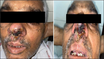
- Necrotic lesion measuring 1.5 × 1 cm on nasal tip over columella extending to left nasal ala, septum, and bilateral vestibule. Overlying skin was erythematous and showed crusting. It was nontender and had no pus discharge or bleeding. Fungal potassium hydroxide (KOH) mount was negative for mucormycosis and was suggestive of acute inflammatory exudate without any granuloma formation.
In the setting of hematological malignancy with AKI, different mechanisms, including tumor lysis syndrome, uric acid nephropathy, obstruction, hypoperfusion, acute tubular necrosis, sepsis, renal vein thrombosis, and direct renal infiltration, were thought of.
Flow cytometry was suggestive of acute promyelocytic leukemia M3 as per French–American–British (FAB) Class. The patient worsened within 48 hours of admission and succumbed to leukostasis. Kidney biopsy showed renal cortex with heavy infiltration of atypical cells within the interstitium and peritubular capillaries [Figure 2a]. These cells showed positive immunoperoxidase staining with myeloperoxidase (MPO) [Figure 2b]. The cause of AKI was thus attributed to renal infiltration from leukemia.

- Kidney biopsy images (a) Light microscopy with periodic acid schiff staining 40×. Several large-sized atypical cells infiltrating in capillary lumina of a glomerulus and (b) Immunocytochemistry. Atypical cells show strong cytoplasmic positivity for myeloperoxidase (MPO). They were negative for CD20, CD3, and CD117 (magnification 40x).
Direct renal infiltration of malignant cells causes AKI in only 1% of leukemia cases.1 Our patient describes a rare presentation of APML with renal infiltration that mimicked sepsis-induced AKI initially. The kidney is the most common extra-hematopoietic and extrareticular organ to be infiltrated by atypical cells.2 Propensity to infiltrate increases with the staging and grading of malignancy. Thirty-three percent patients of AML had infiltration in an autopsy series of 1200 patients.3 Metastatic malignancies can cause cutaneous lesions on the nose as in the index case that resemble a clown’s nose described earlier in case of acute leukemia.4 Because the differential of AKI is broad, kidney biopsy is needed for early diagnosis and special stains can ascertain subtypes of leukemia. Like in our case, atypical cells have strong positivity for MPO.
Declaration of patient consent
The authors certify that they have obtained all appropriate patient consent.
Conflicts of interest
There are no conflicts of interest.
References
- Renal involvement in myeloproliferative and lymphoproliferative disorders. A study of autopsy cases. Gen Diagn Pathol. 1997;142:147-53.
- [PubMed] [Google Scholar]
- Renal lesions associated with malignant lymphomas. Am J Med. 1962;32:184-207.
- [CrossRef] [PubMed] [Google Scholar]
- An Autopsy study of 1206 acute and chronic leukemias (1958 to 1982) Cancer. 1987;60:827-37.
- [CrossRef] [PubMed] [Google Scholar]
- Tumour of the nose as a presenting feature of leukaemia. J Laryngol Otol. 1982;96:83-7.
- [CrossRef] [PubMed] [Google Scholar]


