

Translate this page into:
Radiolucent Calculi in Kidneys on Intravenous Urography: Red Color on Dual-Energy Computed Tomography Clinches the Final Diagnosis
Corresponding author: Akshay Kumar Saxena, Department of Radiodiagnosis and Imaging, Postgraduate Institute of Medical Education and Research (PGIMER), Chandigarh, India. E-mail: fatakshay@yahoo.com
-
Received: ,
Accepted: ,
How to cite this article: Bhatia A, Kotia K, Mavuduru RS, Sodhi KS, Saxena AK. Radiolucent Calculi in Kidneys on Intravenous Urography: Red Color on Dual-Energy Computed Tomography Clinches the Final Diagnosis. Indian J Nephrol. doi: 10.25259/IJN_17_2024
A 1-year-old girl, follow-up case of bilateral hydronephrosis, underwent intravenous urography (IVU) to assess the function and drainage. The plain spot [Figure 1a] of the IVU study showed no radiopaque calculus. Subsequently, in 1-h post-intravenous contrast injection, mild bilateral hydronephrosis was present with two radiolucent filling defects seen, one each in the right and left renal pelvis [Figure 1b] suggestive of radiolucent calculi. Low-dose dual-energy computed tomography (DECT) was subsequently done for further delineation and characterization to decide upon the management. It showed bilateral nephrolithiasis with mild hydronephrosis [Figure 1c and d]. On dual energy evaluation, bilateral renal calculi were red and found to have a uric acid composition [Figure 1e and 1f].
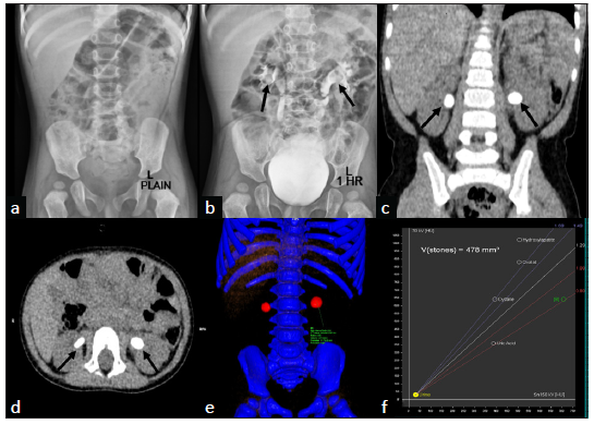
- Plain radiograph (a) shows no radiopaque calculi in the renal fossae on either side, while post-contrast injection 1-h spot (b) shows two radiolucent filling defects in the bilateral renal pelvis (black arrows) with mild hydronephrosis. Coronal (c) and axial (d) multiplanar reformatted images of dual-energy computed tomography (DECT) scan show bilateral renal calculi (black arrows in c and d) with mild hydronephrosis. Color overlay image (e) of DECT scan shows bilateral renal calculi, which are color-coded red. On dual-energy radiograph evaluation (f), left renal calculus (region of interest shown in e) is seen to have a uric acid composition (green circle in f).
Radiolucent stones constitute only 10% of urolithiasis cases. These include uric acid, cysteine, and medication-induced (such as indinavir, triamterene, sulfonamides, and amorphous silica) calculi. DECT can help to differentiate uric acid stones from non-uric acid stones (such as calcium oxalate, calcium phosphate, struvite, cystine, and hydroxyapatite) due to their material decomposition ability.1 Uric acid stones are color-coded red, while the non-uric acid stones are blue. This is important from a management point of view, as the uric acid calculi are medically managed using alkalizing agents. In contrast, non-uric acid stones are treated by lithotripsy or surgery.
Declaration of patient consent
The authors certify that they have obtained all appropriate patient consent.
Conflicts of interest
There are no conflicts of interest.
Reference
- Determination of renal stone composition with dual-energy CT: In vivo analysis and comparison with X-ray diffraction. Radiology. 2010;257:394-401.
- [CrossRef] [PubMed] [Google Scholar]


