Translate this page into:
Adnexal torsion in an adolescent renal transplant recipient
Address for correspondence: Dr. V. V. Mishra, Department of Obstetrics and Gynecology, Institute of Kidney Diseases and Research Center, Dr. HL Trivedi Institute of Transplantation Sciences, Civil Hospital Campus, Asarwa, Ahmedabad, Gujarat, India. E-mail: vvmivf@gmail.com
This is an open access article distributed under the terms of the Creative Commons Attribution-NonCommercial-ShareAlike 3.0 License, which allows others to remix, tweak, and build upon the work non-commercially, as long as the author is credited and the new creations are licensed under the identical terms.
This article was originally published by Medknow Publications & Media Pvt Ltd and was migrated to Scientific Scholar after the change of Publisher.
Sir,
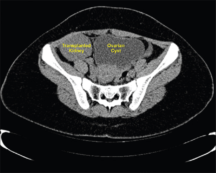
Adnexal torsion is rare in adolescents, with an incidence of 10–12%.[1] Benign adnexal masses (mainly ovarian cysts) have been reported to date in transplanted patients.[2] A 14-year-old girl who had undergone renal transplantation 4 years back presented with acute abdominal pain. Her vital signs were normal. The white blood count was elevated at 14.26 × 1000/cmm, while hemoglobin, hematocrit, and platelets were within normal levels. A basic chemistry panel was normal. Ultrasonography of abdomen and pelvis demonstrated a right enlarged ovary 7 cm × 8 cm × 8 cm with large thin-walled cysts measuring 15 cm × 7 cm × 7 cm with cystic and solid masses and absent color flow. Computed tomography (CT) scan revealed a nonenhancing mixed solid and cystic lesion of size 15 cm × 9 cm seen retroperitoneally extending from L2 to S1 [Figure 1] with associated pelvic free fluid. Uterus and left adnexa normal.

- Hemorrhagic cyst and transplanted kidney
Exploratory laparoscopy showed a 16 cm × 9 cm × 9 cm hemorrhagic cyst extending from right iliac fossa to left hypogastrium with 360° torsion of the right tube, ovary, and cyst. Detorsion was not possible and hence adenexectomy was done. The left ovary was enlarged on naked eye appearance. Histopathology was consistent with hemorrhagic and necrotic adnexa with simple cyst.
The literature review did not reveal any case reports of adnexal torsion in postrenal transplant females. Patients on sirolimus have a high prevalence of benign ovarian cysts.[3] One study reported on the diagnosis of a benign ovarian cyst in 6.7% recipients of the kidney transplant that had large cysts (up to 9 cm) requiring surgical excision.[2] The reported incidence of ovarian cysts in patients on sirolimus plus tacrolimus was higher than that compared to those on tacrolimus plus mycophenolic acid.[3] The hormone profile after kidney transplantation may be impaired; especially the higher levels of estrogens may increase the risk of some gynecological pathology. Thus regular, attentive gynecological care is strongly recommended in female kidney graft recipients.[4] Ovarian torsion with delayed surgical intervention often results in necrosis and loss of ovary. A high index of suspicion and prompt surgical intervention is required in children and adolescent population to salvage the ovary in cases of adnexal torsion. Pre- and post-transplant gynecological evaluation and baseline pelvic ultrasound should be routinely advocated as data suggests there is an increase in the incidence of ovarian cysts in transplant females on immunotherapy.
Financial support and sponsorship
Nil.
Conflicts of interest
There are no conflicts of interest.
References
- A national population-based study of the incidence of adnexal torsion in the Republic of Korea. Int J Gynaecol Obstet. 2015;129:169-70.
- [Google Scholar]
- Unsuspected adnexal masses in renal transplant recipients. J Urol. 1982;128:1017-9.
- [Google Scholar]
- High prevalence of ovarian cysts in premenopausal women receiving sirolimus and tacrolimus after clinical islet transplantation. Eur Soc Organ Transplant. 2009;22:622-5.
- [Google Scholar]
- Function of the ovaries in female kidney transplant recipients. Transplant Proc. 2006;38:180-3.
- [Google Scholar]


