

Translate this page into:
An Uncommon Malignant Suprarenal Mesenchymal Tumor
-
Received: ,
Accepted: ,
How to cite this article: Prachi, Aiyer HM, Sharma G. An Uncommon Malignant Suprarenal Mesenchymal Tumor. Indian J Nephrol. 2024;34:204. doi: 10.4103/ijn.ijn_278_23
Dear Editor,
Primary adrenal leiomyosarcoma is a rare malignant tumor. Metastatic adrenal leiomyosarcoma and extension from the retroperitoneum are more frequent. Leiomyosarcoma occurs primarily in the myometrium, retroperitoneum, and soft tissues of the extremities. Herein we report a case in a 31 year old female who presented with abdominal pain. Contrast-enhanced computed tomography (CECT) and contrast-enhanced magnetic resonance imaging (CEMRI) abdomen revealed a large heterogeneously enhancing mass in the right suprarenal region abutting the surrounding structures such as liver, superior pole of right kidney, adjacent inferior vena cava, second part of duodenum, and right renal vein, causing its upliftment [Figure 1a]. Further, she underwent resection of the mass along with right nephrectomy. Pathological findings revealed a nonencapsulated mass measuring 8.5 × 5.5 × 4.5 cm. Cut surface appeared fleshy with hemorrhagic and necrotic areas [Figure 1b]. Microscopically, solid pattern of spindle cells showing marked pleomorphism with 30% areas of necrosis and 10–12 muscle fibers/10 high power fields, with compressed adrenal gland at the periphery was seen [Figure 1c]. Immunohistochemically, intense positivity for desmin, smooth muscle actin [Figure 1d], caldesmon, and vimentin was observed. Considering the aforementioned features, a diagnosis of conventional primary leiomyosarcoma of the adrenal gland, FNCLCC Grade 2 was rendered. After 15 months of follow-up, the patient remained free of recurrence. Adrenal leiomyosarcomas are rare tumors, usually diagnosed at an advanced stage due to nonspecificity of symptoms, contributing to poor prognosis. The gold standard for treatment is surgical excision, followed by chemotherapy and radiotherapy.
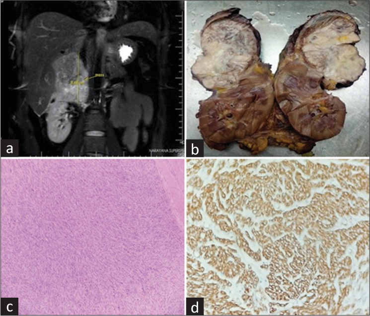
- Images of Primary adrenal leiomyosarcoma. (a) Imaging showing a large heterogeneously enhancing mass in the right suprarenal region abutting the surrounding structures. (b) Gross photograph of right nephrectomy specimen with a well defined fleshy suprarenal mass. (c) Microphotograph showing solid pattern of spindle cells showing marked pleomorphism and focal necrosis (H and E X100). (d) Immunoreactivity for Smooth Muscle actin (IHC stain for SMA X400).
Declaration of patient consent
The authors certify that they have obtained all appropriate patient consent.
Conflicts of interest
There are no conflicts of interest.
Financial support and sponsorship
Nil.


