Translate this page into:
An unusual cause of lower limb ulcer in renal allograft recipient
Address for correspondence: Dr. N. Raveendran, Department of Nephrology, SMS Medical College, Jaipur - 302 004, Rajasthan, India. E-mail: nishadravi@gmail.com
This is an open access article distributed under the terms of the Creative Commons Attribution-NonCommercial-ShareAlike 3.0 License, which allows others to remix, tweak, and build upon the work non-commercially, as long as the author is credited and the new creations are licensed under the identical terms.
This article was originally published by Medknow Publications & Media Pvt Ltd and was migrated to Scientific Scholar after the change of Publisher.
Sir,
Fungal infection following solid-organ transplantation remains a major cause of morbidity and mortality. Cryptococcosis is the third most common invasive fungal infection in organ transplant recipients, after candidiasis and aspergillosis.[1] The incidence of the infection appears to vary between 0.8% and 5.8% depending upon the type and intensity of immunosuppression used.[2] Cryptococcus neoformans has been shown to cause almost every type of cutaneous lesion. The lesions may occur as ulcers, pustules, granulomata, abscesses, and herpetiform or molluscum contagiosum-like lesions. In this communication, we describe a case of cutaneous cryptococcosis in a renal transplant recipient manifesting as cellulitis, which was the only sign of the disease when diagnosis was made.
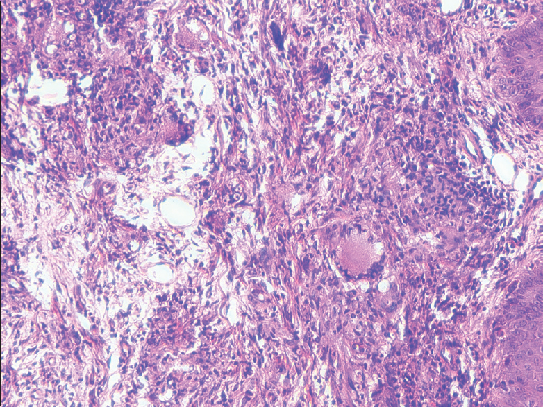
A 45-year-old male who underwent living-related donor renal transplant 8 years back with sister as donor presented to us with complaints of nonhealing ulcer on the right thigh for 1 month. Following transplantation, he was maintained on triple immunosuppressants (prednisolone, tacrolimus, and mycophenolate mofetil). First 3 years of his posttransplant period were uneventful, following which he developed gradual rise in serum creatinine and the serum creatinine remained more or less at a constant level of 3.5 mg/dl. Renal allograft biopsy was suggestive of features of chronic antibody-mediated rejection with 30–40% interstitial fibrosis and tubular atrophy. Except for the raised creatinine levels, the patient was relatively asymptomatic for the past 4 years, until he developed an ulcer on his right thigh. It started in the form of a pustule with surrounding edema and pain, which got ruptured later to become an oval shaped ulcer of approximately 10 cm × 8 cm in size. There was no history of any trauma at the site, no history of any insect bite, and no history of any fever. On local examination, an ulcer of 10 cm × 8 cm was present on the right thigh with irregular margins, slopy edges, floor filled with slough, and base probably subcutaneous tissue [Figure 1]. There was surrounding erythema and the right inguinal lymph nodes were enlarged. His systemic examination was otherwise normal. His routine investigations were normal except for raised blood urea and serum creatinine. High-resolution computed tomography thorax was normal. Cryptococcal antigen test was positive by latex agglutination. Biopsy of the lesion revealed entire dermis infiltrated with epitheloid cells granuloma with foreign body giant cells. Numerous cryptcocci were seen within the macrophages and giant cells [Figure 2]. The yeast bodies stained deeply with periodic acid–Schiff stain. The patient was started on injection amphotericin B deoxycholate 0.1 mg/kg for 14 days followed by tablet itraconazole 200 mg thrice daily for 45 days. At the end of 1 month of therapy, there was complete healing of the lesion while cryptococcal antigen test was negative. Two months after completion of therapy, cryptococcal antigen test continued to be negative.

- Image showing lower limb ulcer

- Skin biopsy showing cryptococcal granuloma
The immunosuppressive regimens used in solid organ transplantation are potent inhibitors of T-cell immunity. This predisposes patients to infection by pathogens such as C. neoformans. Our patient was on triple immunosuppression including prednisolone. Usage of corticosteroids has been found to be the most common risk factor in an analysis of 37 patients with primary cutaneous cryptococcosis.[3] Although a rarity, several cases of cellulitis have been reported, which appear to be mainly restricted to lower parts of the body, especially the legs, as was seen in the present. The laboratory diagnosis of cryptococcosis is established by the isolation of the organism in culture, histopathology, or detection of its capsular polysaccharide antigen.[4] In conclusion, cellulitis is an unusual presentation of cryptococcal infection in renal allograft recipients. Patients are often treated with antibiotics before a definitive diagnosis is made, delaying appropriate therapy. Hence, a high clinical suspicion and early initiation of therapy are needed to recognize and treat patients effectively.
Financial support and sponsorship
Nil.
Conflicts of interest
There are no conflicts of interest.
References
- Early-onset of disseminated cryptococcal infection in two renal transplant recipients. Clin Nephrol. 2011;75:542-6.
- [Google Scholar]
- Fungal infection following renal transplantation. Arch Intern Med. 1975;135:1163-72.
- [Google Scholar]
- Primary cutaneous cryptococcosis in immunocompetent and immunocompromised hosts. Med Mycol. 2003;41:177-88.
- [Google Scholar]
- Cryptococcal neoformans profiles in peripheral blood neutrophils: An unusual presentation. Indian J Pathol Microbiol. 2008;51:296-7.
- [Google Scholar]


