

Translate this page into:
Bedside Method to Check the Adequacy of Kidney Biopsy Sample with a Smartphone Camera and Macro Lenses: A Prospective Cohort Study
Corresponding author: Dr. Suny S. Modi, Department of Nephrology, Vishesh - Jupiter Hospital, Ring Road, Near Teen Imli Square Indore, Madhya Pradesh, India. E-mail: sunynephrology@gmail.com
-
Received: ,
Accepted: ,
How to cite this article: Modi SS, Ramamurthy S, Balasubramanian S, Kumar S, Daruwala F. Bedside Method to Check the Adequacy of Kidney Biopsy Sample with a Smartphone Camera and Macro Lenses: A Prospective Cohort Study. Indian J Nephrol. 2024;34:233–6. doi: 10.4103/ijn.ijn_323_23
Abstract
Background:
The utilization of smartphone-assisted evaluation is emerging in the field of histopathology. This technique improves the adequacy of samples at the bedside, avoids procedure-related complications, reduces unnecessary repeat biopsies, and saves the cost of the procedure. This study aims to compare the number of glomeruli in a renal biopsy specimen obtained by an ultrasound-guided percutaneous needle biopsy, counted at the bedside using a smartphone fitted with a 16-megapixel macro lens (Bedside method) with that observed under a light microscope after the processing of the biopsy specimen (LM method).
Materials and Methods:
In this prospective cohort study, 24 consecutive adult patients (48 kidney biopsy samples) who underwent kidney biopsies were enrolled. All specimens were extracted by an ultrasound-guided percutaneous renal biopsy from the lower pole of the left kidney. Patients’ demographics and clinical data were prospectively collected. The number of glomeruli in all the biopsy specimens was counted using a smartphone fitted with a 16-megapixel macro lens at the bedside (Bedside method) and subsequently under a light microscope by a pathologist after processing the biopsy specimen (LM method). Seven or more glomeruli in the specimen were considered adequate in our study.
Results:
The mean age of patients at biopsy was 46.9 ± 16 years with slightly male predominance (54.2%). A total of 47 specimens were obtained from 24 patients. Of the 24 patients, 22 had native kidney biopsy and 2 had renal allograft biopsy. The average number of cores obtained per patient was 1.96. The length of core specimens ranged from 1.5 to 2 cm. A good agreement was found between bedside adequacy and slide adequacy, κ =0.684, P = 0.000. The positive agreement rate and negative agreement rate were 91.4% and 23.1%, respectively.
Conclusion:
In the modern era of technology, the smartphone is a good tool to evaluate the adequacy of biopsy specimens at the bedside.
Keywords
Adequacy
biopsies
immunofluorescence
kidney
light microscopy
smartphone


Introduction
Percutaneous kidney biopsy (PKB) is regarded as the gold standard investigation in diagnosing native as well as renal allograft diseases.1 The quality of sample size for tissue diagnosis has improved dramatically, as the PKB has evolved from blind aspiration technique to the use of an ultrasound-guided spring-loaded automated kidney biopsy gun with reduced complication rates.2 Since the inception of PKB, controversy still surrounds the definition of an adequate kidney biopsy specimen in order to achieve diagnostic accuracy. The minimum sample size required for diagnosis varies greatly with different renal pathologies. Five glomeruli would be considered to be adequate to diagnose most glomerular diseases, six to ten for tubulointerstitial disease, and seven for transplanted kidneys.1 Of the various glomerular diseases, membranous nephropathy can be diagnosed from a single glomerulus, but a minimum of 25 glomeruli may be required for a 95% chance of diagnosing a focal and segmental glomerulosclerosis.3,4
Typically, glomeruli in the cortex appear as reddish, dotted structures, and the medulla appears reddish streaks lacking typical glomerular dotted appearance.2 Ideally, the first step in the evaluation of a kidney biopsy specimen is a bedside wet mount preparation of the tissue sample for adequacy, followed by allocation of tissue for LM, immunofluorescence (IF), and electron microscopy at the same time.5,6 This method has the benefits of checking the adequacy of specimens with a proper allocation of specimens. Most institutions do not follow this bedside evaluation procedure as it is more cumbersome and needs a trained specialist for the same.2 The rate of inadequate sampling increased from 12.5% to 21.6% after abandoning bedside microscopic evaluation.4 Another study demonstrated that the likelihood of getting inadequate specimens was nearly four times greater without on-site evaluation than with on-site evaluation.7 With significant breakthroughs in modern technology, more recently, the use of smartphone cameras has been evolving for bedside evaluation of kidney biopsy. Although some endeavours have been made to address this novel smartphone-assisted approach, few studies have been published in India. Our study aims to count the number of glomeruli from PKB using a smartphone fitted with a 16-megapixel macro lens at the bedside (Bedside method, BS) and subsequently under a light microscope by a pathologist after processing of the biopsy specimen (Light microscopy method, LM).
Materials and Methods
This prospective cohort study comprised 24 consecutive adult patients (47 kidney biopsy samples) who underwent kidney biopsies in our tertiary health care center from June 2017 until January 2018. The Institutional ethical approval has been obtained (Approval No: RES-1319-17). The study was conducted in accordance with the Declaration of Helsinki Guidelines, and written consent was obtained from all participants in the study.
Patient demographics (age, gender) and clinical data (left kidney size, kidney echoes, serum creatinine, estimated glomerular infiltration rate (eGFR) before biopsy, and biopsy details) were collected. All biopsy specimens were extracted from the lower pole of the left kidney and evaluated by Nephrology fellows with the assistance of a radiologist under real-time ultrasound imaging guidance. According to our institutional protocol, semiautomated spring-loaded core biopsy needles were used to perform all kidney biopsies. The length of each core specimen was measured immediately on a moistened, nonadhesive dressing pad.
We used a Samsung 16-megapixel camera mobile fitted with a of 5 × Macro Photo Lens (Black). For this research, photographs of the specimens were obtained with the help of the smartphone, which was held approximately 3 inches away from the specimens while capturing pictures. Autofocus was used without using optical zoom.
The renal capsule looked like a fibrofatty tissue string; fat was yellowish. Renal medulla had reddish brown speckles on the background of light tan tissue [Figure 1], whereas the cortex had a paler appearance and the glomeruli appeared as reddish dots [Figure 2]. Subsequently, the biopsy specimens were sent to the pathology department, where they were processed for slide preparation and subjected to LM/IF. Microscopic evaluation was performed by an expert pathologist. We defined the adequacy of specimens as more than 7 glomeruli in a specimen. Once adequate specimens were obtained, the procedure was completed and further statistical analysis was done. We compared the number of glomeruli counted by the Bedside method and by LM method.
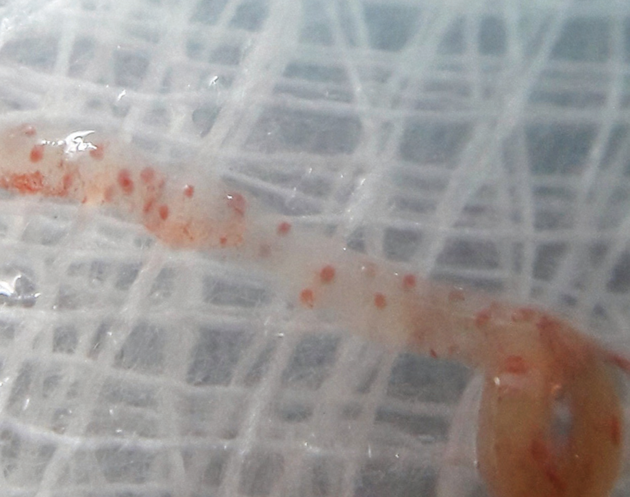
- Cortical tissue with glomeruli appears as reddish, circular structures.
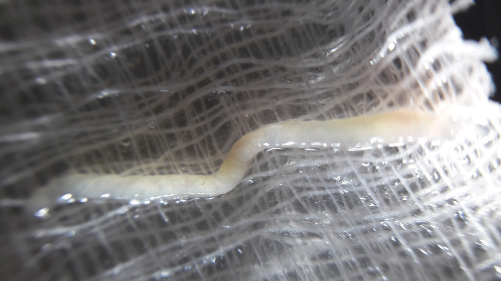
- Predominant medullar tissue-lacking typical glomerular dotted appearance.
Statistical analysis was performed using commercial software packages (IBM SPSS Statistics for Windows, version 19.0, IBM Corp., Armonk, NY, USA). Continuous variables were expressed as mean and standard deviation or range, while categorical variables were presented as frequency and percentages. Cohen’s Kappa statistic was used to assess the reliability of rates between two diagnostic measures.
Results
Forty-eight kidney biopsy samples (24 consecutive subjects) were analyzed. In this study cohort, the mean age at biopsy was 46.9 ± 16 years with slightly male predominance (54.2%). The mean serum creatinine and eGFR were 3.2 ± 3.3 mg/dl and 46.6 ± 40.1 mL/min/1.73m2, respectively. The most common biopsy indication was azotemia (66%), followed by proteinuria (45%) and nonproteinuric kidney disease (55%). The baseline demographic and clinical characteristics of the study population are outlined in Table 1.
| Characteristics | Total patients (n = 24) |
|---|---|
| Age at biopsy, years | 46.9±16.3 |
| Male | 13 (54.2%) |
| Left kidney size, cm | 10.3 ± 1.3 |
| Kidney echoes | |
| Normal | 12 (50%) |
| Increased with CMD | 12 (50%) |
| Serum creatinine (mg/dl) | 3.2 ± 3.3 |
| eGFR before biopsy (mL/min/1.73 m2) | 46.6 ± 40.1 |
| Indication, n (%) | |
| Azotemia | 16 (66%) |
| Proteinuric Kidney disease | 11 (45%) |
| Nonproteinuric Kidney disease | 13 (55%) |
Data are expressed as mean ± standard deviation and counts. CMD, Renal corticomedullary differentiation; eGFR, estimated glomerular filtration rate.
Biopsy details of the study population are presented in Table 2. Of the total 24 patients, 22 had native and 2 had renal allograft biopsies. The number of cores obtained per patient was 1.96. The length of core specimens ranged from 1.5 to 2 cm.
| Characteristics | Value |
|---|---|
| Number of biopsies | 24 |
| Biopsy type, n (%) | |
| Native kidney biopsy | 23/24 (95.8%) |
| Transplant biopsy | 1/24 (4.2%) |
| Biopsy needle, n (%) | |
| 18G | 6/24 (25%) |
| 16G | 18/24 (75%) |
| Total number of cores | 47 |
| Number of cores per patient | 1.96 |
| Mean length of core (cm), range | 1.5–2 |
Data are expressed as counts and range
Thirty-five (72.9%) patients had adequate and 13 (27.1%) patients had an inadequate number of glomeruli by Bedside method, while 35 (72.9%) had adequate and 13 (27.1%) had an inadequate number of glomeruli in the assessment done by LM/IF method.
We compared the agreement between both the methods. A good agreement was found between bedside adequacy and slide adequacy, κ = 0.684, and P = 0.000 [Table 3].
| Comparison between bedside adequacy with slide adequacy | ||
|---|---|---|
| Bedside adequacy | Slide adequacy | n (%) |
| Adequate | Adequate | 32 (66.7%) |
| Inadequate | Inadequate | 10 (20.8%) |
| Adequate | Inadequate | 3 (6.3%) |
| Inadequate | Adequate | 3 (6.3%) |
| Positive agreement % | 91.4% | |
| Negative agreement % | 23.1% | |
| Overall agreement % | 72.9% | |
| Cohen’s kappa statistics | 0.684 | |
| P value | 0.000 | |
Data are expressed as counts. P < 0.05 was considered as statistically significant
The final pathology outcomes of our study population are demonstrated in Table 4. No complications occurred following the procedure, and recovery for all patients was uneventful.
| Outcomes | Total patients (n=24) |
|---|---|
| Cortical tissue | |
| Able to make diagnosis | 24 (100%) |
| Diagnosis | |
| Membranous nephropathy with FSGS | 1 (4.2%) |
| Diabetic nephropathy | 5 (20.8%) |
| Acute cellular rejection (ACR) and diabetic nephropathy | 1 (4.2%) |
| Lupus along with diabetic nephropathy | 1 (4.2%) |
| Chronic diabetic nephropathy along with acute tubular necrosis | 1 (4.2%) |
| IgA nephropathy | 3 (8.3%) |
| Lupus nephritis | 1 (4.2%) |
| Acute interstitial nephritis | 1 (4.2%) |
| Immunoglobulin G4-related disease | 1 (4.2%) |
| Acute rejections | 1 (4.2%) |
| Diabetic nephropathy with acute phosphate nephropathy | 1 (4.2%) |
| Class V lupus nephritis | 4 (16.7) |
| FSGS | 1 (4.2%) |
| Crescentic glomerulonephritis | 1 (4.2%) |
| Minimal change disease | 1 (4.2%) |
Data are expressed as mean±standard deviation and counts. FSGS, Focal segmental glomerulosclerosis
Discussion
The smartphone camera-assisted approach is a novel approach that is easy to adopt since it is simple, cost-effective, and easily reproducible. Sirithanaphol reported a transplant biopsy inadequacy rate of 7% using a smartphone-based magnifying device than 21.3% without it.7 Another prospective cohort study consisting of 57 kidney biopsy cores (20 patients) conducted by Singh et al.8 compared bedside evaluation of specimens with evaluation done by a pathology technician using a dissecting microscope, which showed an excellent agreement between smartphone-assisted bedside evaluation and light microscopic evaluation (Cohen’s kappa, 0.83; positive adequacy rate, 97.4%; negative adequacy rate, 83.3%).6 Our study showed a good agreement of kidney biopsy sample adequacy by smartphone-assisted bedside evaluation with light microscopic slide evaluation. Despite the success demonstrated, there is a discrepancy in the study results (Cohen’s kappa, 0.684; positive adequacy rate, 91.4%; negative adequacy rate, 23.1%) from the findings reported by Singh et al.,8 which could be attributed to the quality of the smartphone camera.
Currently, most kidney biopsies are performed by nephrologists, who frequently lack access to an on-site laboratory facility for the evaluation of the adequacy of the biopsy specimen. In such circumstances, the use of smartphone-assisted approach could be beneficial to assess adequacy of the specimens at the bedside, thus reducing the requirement for repeat biopsies, especially in high-risk patients which, in turn, prevent complications and lower the overall cost of care.9
The key strength of our study was that the pathologists were completely blinded for the study which mitigated the potential for bias on evaluation of slide specimens. However, a major drawback associated with this study is perception bias – nephrologists are more likely to consider a specimen adequate in patients at a high risk of bleeding or needing several needle passes. In the end, this study provides a solid foundation on which future multicenter studies with larger sample size and more data on transplanted kidney biopsy and head-to-head comparison, before generalization can build.
Conclusion
This study demonstrates that smartphone is a promising tool for assessing bedside adequacy of renal biopsy specimens in resource-constrained healthcare systems. It is a simple and an easy technique that does not need any expert training for observation.
Conflicts of interest
There are no conflicts of interest.
References
- Basics of kidney biopsy: A nephrologist's perspective. Indian J Nephrol. 2013;23:243-52.
- [CrossRef] [PubMed] [Google Scholar]
- The impact of renal tissue procurement at bedside on specimen adequacy and best practices. Am J Clin Pathol. 2019;151:205-8.
- [CrossRef] [PubMed] [Google Scholar]
- Ad hoc committee on renal biopsy guidelines of the renal pathology society. Practice guidelines for the renal biopsy. Mod Pathol. 2004;17:1555-63.
- [CrossRef] [PubMed] [Google Scholar]
- Role of on-site microscopic evaluation of kidney biopsy for adequacy and allocation of glomeruli: Comparison of renal biopsies with and without on-site microscopic evaluation. Pathologica. 2013;105:342-5.
- [PubMed] [Google Scholar]
- Improvement of allograft kidney biopsy yield by using a handheld smartphone microscope as an on-site evaluation device. Heliyon. 2021;7:e07189.
- [CrossRef] [PubMed] [Google Scholar]
- Use of a smartphone camera at the bedside to assess adequacy of kidney biopsies. J Am Soc Nephrol. 2021;32:3024-6.
- [CrossRef] [PubMed] [Google Scholar]
- Kidney biopsy adequacy: A metric-based study. Am J Surg Pathol. 2019;43:84-92.
- [CrossRef] [PubMed] [Google Scholar]


