Translate this page into:
Carbamylated Hemoglobin can Differentiate Acute Kidney Injury from Chronic Kidney Disease
This is an open access journal, and articles are distributed under the terms of the Creative Commons Attribution-NonCommercial-ShareAlike 4.0 License, which allows others to remix, tweak, and build upon the work non-commercially, as long as appropriate credit is given and the new creations are licensed under the identical terms.
This article was originally published by Medknow Publications & Media Pvt Ltd and was migrated to Scientific Scholar after the change of Publisher.
Abstract
Carbamylated hemoglobin (CarHb) was found to have a potential role in the differentiation of patients with acute kidney injury (AKI) from chronic kidney disease (CKD). This study was aimed at the evaluation of the diagnostic performance and usefulness of CarHb in the differentiation of AKI from CKD. Forty patients with renal disease and twenty age- and sex-matched healthy controls were included in the study. Urea, creatinine, Hb, and CarHb were measured in all the subjects. Patients with AKI and CKD were found to have significantly increased levels of CarHb when compared to controls (P < 0.05 for both groups). Patients with CKD had significantly increased levels of CarHb when compared to patients with AKI (P < 0.05). CarHb showed significant positive correlation with urea in patients with renal disease (r = 0.776, P < 0.0001). Significant area under curve (AUC = 0.840, P < 0.0001) was obtained for CarHb and a cut-off value of 98.33 μg VH/g Hb resulted with the best combination of 85% sensitivity and 75% specificity. CarHb may provide clinical utility since patients with AKI and CKD have similar clinical presentation usually. A cut-off value of 98.33 μg VH/g Hb has been found to be useful to differentiate AKI from CKDs.
Keywords
Acute kidney injury
carbamylated hemoglobin
chronic kidney disease
Introduction
Assessment of renal function is one of the most important steps in acute emergencies to identify renal failure with either acute kidney injury (AKI) or chronic kidney disease (CKD). AKI is defined as abrupt (i.e., within 48 h) reduction in kidney function, currently defined as an absolute increase in serum creatinine level more than or equal to 0.3 mg/dl (≥ 26.5 μmol/L). AKI represents a substantial risk factor for CKD.[12] CKD is characterized by a progressive and ongoing loss of kidney function of not <3 months duration with or without decrease in glomerular filtration rate (GFR).[3]
Carbamylated hemoglobin (CarHb), which is formed as a result of the reaction of isocyanate with N-terminal valine residues of the α and β chains of Hb is found in high levels in patients with renal failure.[4] Under physiological conditions, the level of isocyanate is approximately 1% that of urea.[5]
Evaluation of CarHb in patients with various degrees of renal function found that measurement of CarHb provides potential clinical value and has been suggested to be useful in differentiating patients with AKI from those with CKD.[67] Based on its importance, this study was aimed to evaluate the diagnostic performance of CarHb in the diagnosis and differentiation of AKI and CKD.
Material and Methods
Forty patients with either AKI (20 patients) or CKD (20 patients) as per Kidney Disease Improving Global Outcomes guidelines attending nephrology outpatient department and aged 20–65 years were enrolled in this study and compared with age- and sex-matched healthy controls. All the subjects were categorized into three groups as Group 1 (controls, n = 20), Group 2 (patients with AKI, n = 20), and Group 3 (patients with CKD, n = 20). Patients with acute or chronic infections, malignancy, pregnancy, and lactation, belonging to pediatric age group, patients with acute on chronic kidney disease and subjects not willing to participate in the study were excluded from the study. Informed consent was obtained from all the subjects and the study was approved by the Institutional ethics committee.
A volume of 5mL of venous blood was collected from all the subjects into plain tubes and ethylenediaminetetraacetic acid tubes. Blood collected in plain tubes was allowed to clot, serum separated by centrifugation at 2000 rpm for 15 minutes. Hemolysate was prepared from the whole blood by washing the erythrocytes 4 times with saline before lysing with equal volumes of cold distilled water. The separated serum and hemolysate were transferred into appropriately labeled aliquots and stored at −80°C until further analysis. Hb was estimated by cyanmethemoglobin method, and CarHb was measured using Modified Rosen's colorimetric method.[89] Measurement of urea was done by enzymatic timed fixed endpoint method, and creatinine was assayed using Modified Jaffe's rate kinetic method on Beckman unicel DXC 600 analyzer, Beckman, USA.
Statistical analysis
Continuous variables were expressed as mean ± standard deviation. Comparison of variables between the three study groups was made using Kruskal–Wallis test, followed by pairwise comparisons. The association between CarHb and study parameters was assessed using Spearman's rank correlation analysis. The utility of CarHb in differentiating AKI from CKD was determined by constructing receiver operating characteristic (ROC) curves. A P < 0.05 was considered statistically significant. All the analyses were performed using Microsoft excel spread sheets and MedCalc version 12.2.1 statistical software (Broekstraat, Mariakerke, Belgium).
Results
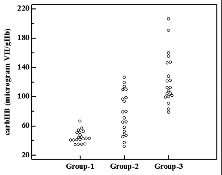
The demographic characteristics and biochemical parameters studied in controls, patients with AKI and patients with CKD are shown in Table 1. The scatter plot of CarHb levels in the groups studied is presented in Figure 1. Serum urea and creatinine were found to be significantly higher in both groups of patients with renal disease than controls. Patients with CKD had significantly higher levels than in patients with AKI. Similarly, CarHb levels were significantly higher in patients with AKI as well as CKD than controls. The levels were found to be highest in patients with CKD.


- Carbamylated hemoglobin levels in controls, patients with acute kidney injury and patients with chronic kidney disease. Group-1=controls; Group-2=patients with acute kidney injury; Group-3=patients with chronic kidney disease
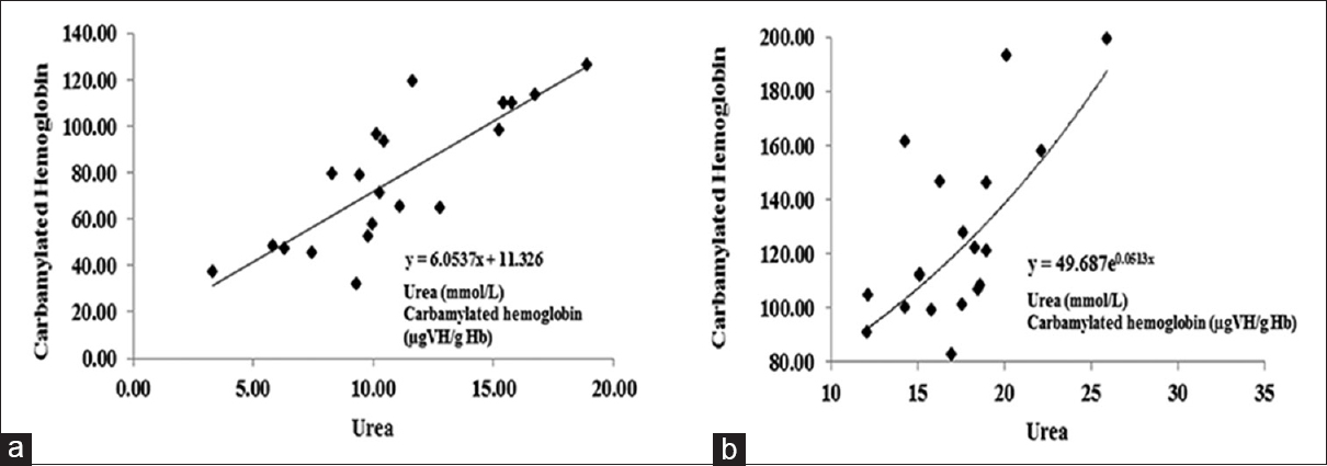
When the association of CarHb with the study parameters was analyzed, CarHb showed significant positive correlation with urea (r = 0.776, P < 0.0001) in all patients with renal disease. However, no correlation was observed between CarHb and creatinine (r = 0.157, P = 0.333) or Hb (r = 0.126, P = 0.439). Figure 2 shows the relationship between CarHb and urea in AKI patients [Figure 2a] and CKD patients [Figure 2b].

- Regression curve analysis between urea and carbamylated hemoglobin levels in acute kidney injury patients (a) and chronic kidney disease patients (b)
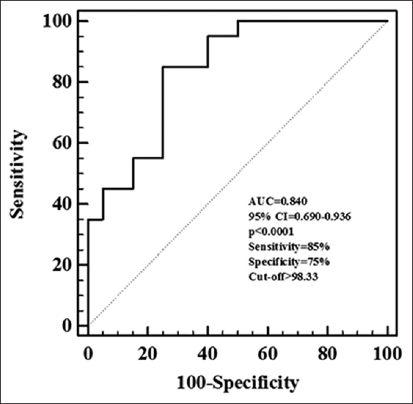
ROC curve analysis performed to study the utility of CarHb in the differentiation of AKI from CKD showed significant area under curve (AUC = 0.840, 95% confidence interval [CI] = 0.690–0.936, P < 0.0001). A cut-off value of 98.33 μg VH/g Hb was obtained with the best combination of 85% sensitivity and 75% specificity to differentiate AKI from CKD [Figure 3].

- Receiver operating characteristic curve analysis of carbamylated hemoglobin. Receiver Operating Characteristic curve analysis done to study the utility of carbamylated hemoglobin in the differentiation of acute from chronic kidney disease showed significant area under curve (AUC=0.840, 95% CI=0.690-0.936, p<0.0001). A cut-off value of 98.33 μgVH/g Hb was obtained with the best combination of 85% sensitivity and 75% specificity to differentiate AKI from CKD
Discussion
Accumulation of nitrogenous waste products occurs as an early sign observed in patients with renal dysfunction and often occurs before the appearance of other symptoms. Urea is one of the first nitrogenous waste substances that accumulate in the blood in renal disease and urea levels increase progressively with worsening renal dysfunction. Hence, assessment of renal function is usually done by the measurement of urea and creatinine levels in serum or plasma and calculation of GFR using prediction equations.
Carbamylation is a nonenzymatic posttranslational modification of proteins involving the reaction of isocyanic acid with the functional groups of amino acids and proteins. Isocyanic acid, which is the protonated and reactive form of cyanate is formed as a spontaneous dissociation product of urea and reacts with the α and ε amino groups of several proteins, including Hb. Carbamylation of Hb is thus an example of nonenzymatic posttranslational modification of proteins and is one of the widely studied reactions and is also found to contribute to uremic toxicity.[1011] In this study, CarHb levels were measured in patients with AKI and CKD and compared with those of healthy controls [Table 1]. It was found that both groups of patients with renal failure had significantly higher levels of CarHb as compared to controls (P< 0.05 for both groups). These findings are similar to those reported by Wynckel et al.,[4] in their study. Under physiological conditions, the concentration of isocyanate is about 1% that of urea. In conditions of renal failure, retention of urea along with other metabolites occurs, and the constant exposure of Hb to increased urea levels results in the increased levels of CarHb.
Patients with CKD in the present study had significantly higher levels of CarHb than those in patients with AKI (P < 0.05). Earlier studies have also reported similar findings. A study by Wynckel et al.,[4] showed that patients with chronic renal failure (CRF) had higher CarHb levels than patients with acute renal failure (ARF). Similarly, Stim et al.,[12] observed that mean CarHb levels were highest in patients with CRF, intermediate in end-stage renal disease on hemodialysis and lowest in ARF, when compared to controls.
Measurement of protein carbamylation was found to reflect a time-averaged record of urea concentration, thus helping in the characterization of duration and severity of renal disease.[9] Hence, CarHb is considered as a potential marker of chronic urea exposure as the rate of its formation is influenced by mean blood urea concentration and the length of exposure to urea.[12] The higher the duration of exposure of the proteins to high urea concentration, the higher is the amount of CarHb that is formed.[4] In this context, Stim et al.,[12] observed a linear relationship between blood urea nitrogen and CarHb in ARF; in the case of CRF patients, the relationship was found to be exponential. Thus, as the concentration of urea increases, patients with CKD manifest an exponential increase in the carbamylation of Hb. Moreover, it has also been reported that in addition to blood urea concentration and duration of exposure to urea, other factors intrinsic to CRF also contribute to the carbamylation rate.[411] All these factors might have contributed to the higher CarHb levels observed in CKD patients when compared to patients with AKI in the present study. It was found that CarHb was significantly correlated with urea (r = 0.776, P < 0.0001) in all patients with renal failure in the present study. However, no correlation was observed between CarHb and creatinine (r = 0.157, P = 0.333). These findings are in accordance with earlier reports.[12]
Based on the differences observed in the carbHb levels between patients with AKI and CKD in the present study, ROC curve was constructed to assess the clinical usefulness of CarHb in the differentiation of AKI from CKD. A significant AUC was obtained (AUC = 0.840, 95% CI = 0.690–0.936, P < 0.0001) and a cut-off value of 98.33 μg VH/g Hb resulted in a sensitivity and specificity of 85% and 75%, respectively. In this context, Wynckel et al.,[4] obtained a cut-off value of 100 μg CV/g Hb with a sensitivity of 94% and specificity of 92%. These findings indicate that CarHb may be used to differentiate patients with potentially reversible renal disease from CKD.
Study with AKI and CKD groups with matched urea concentration would have been better so that only duration of increase would have been the major differentiating factor. In spite of this limitation to obtain urea matched AKI and CKD patients, the findings of the present study support earlier observations.
Conclusion
Hence, CarHb can be used to differentiate patients with AKI from CKD. A cut-off value of 98.33 μg VH/g Hb has been found to be useful in differentiating patients with AKI from those with CKD. This may provide clinical utility since patients with AKI and CKD usually have a similar clinical presentation and with different management strategies.
Financial support and sponsorship
Nil.
Conflicts of interest
There are no conflicts of interest.
References
- Chronic kidney disease after acute kidney injury: A systematic review and meta-analysis. Kidney Int. 2012;81:442-8.
- [Google Scholar]
- The risk of acute renal failure in patients with chronic kidney disease. Kidney Int. 2008;74:101-7.
- [Google Scholar]
- K/DOQI clinical practice guidelines for chronic kidney disease: Evaluation, classification, and stratification. Am J Kidney Dis. 2002;39(2 Suppl 1):S1-266.
- [Google Scholar]
- Kinetics of carbamylated haemoglobin in acute renal failure. Nephrol Dial Transplant. 2000;15:1183-8.
- [Google Scholar]
- Reactions of the cyanate present in aqueous urea with amino acids and proteins. J Biol Chem. 1960;235:3177-81.
- [Google Scholar]
- Protein carbamylation in kidney disease: Pathogenesis and clinical implications. Am J Kidney Dis. 2014;64:793-803.
- [Google Scholar]
- Carbamylation-derived products: Bioactive compounds and potential biomarkers in chronic renal failure and atherosclerosis. Clin Chem. 2011;57:1499-505.
- [Google Scholar]
- Carbamylated haemoglobin determination in assessment of renal disease. Arch Biochem Biophys. 1957;67:10-5.
- [Google Scholar]
- A colorimetric method for measurement of carbamylated haemoglobin in patients with chronic kidney disease using a spectrophotometer. J Med Med Sci. 2012;3:494-8.
- [Google Scholar]
- Protein carbamylation links inflammation, smoking, uremia and atherogenesis. Nat Med. 2007;13:1176-84.
- [Google Scholar]
- Chronic increase of urea leads to carbamylated proteins accumulation in tissues in a mouse model of CKD. PLoS One. 2013;8:e82506.
- [Google Scholar]
- Factors determining hemoglobin carbamylation in renal failure. Kidney Int. 1995;48:1605-10.
- [Google Scholar]


