

Translate this page into:
Diphallia with Duplicated Urethra and Urinary Bladder: A Rare Congenital Genitourinary Tract Anomaly
 , Anmol Bhatia1,, Nitin James Peters2, Akshay Kumar Saxena1, Kushaljit Singh Sodhi1,3
Corresponding author: Anmol Bhatia, Department of Radiodiagnosis and Imaging, Postgraduate Institute of Medical Education and Research, Chandigarh, India. E-mail: anmol_bhatia26@yahoo.co.in
-
Received: ,
Accepted: ,
How to cite this article: Hegde SG, Bhatia A, Peters NJ, Saxena AK, Sodhi KS. Diphallia with Duplicated Urethra and Urinary Bladder: A Rare Congenital Genitourinary Tract Anomaly. Indian J Nephrol. 2025;35:306-7. doi: 10.25259/IJN_499_2024
A 5-month-old boy presented to the outpatient department with complaints of two separate phalluses [Figure 1a]. On examination, the upper phallus was smaller and had normal spontaneous urine excretion, while the lower phallus was larger and exhibited occasional urine excretion. A retrograde urethrogram study using an infant feeding tube (IFT) revealed opacification of two distinct and separate penile and bulbar urethras, each showing a normal course, caliber, and outline. Subsequently, when the IFT was gently advanced across the right and left posterior urethra sequentially, it revealed opacification of two small, non-communicating contrast-filled structures, suggesting complete urinary bladder duplication. Additionally, there was reflux of contrast into the left ureter and pelvicalyceal system from the left-sided urinary bladder [Figure 1b-1e]. Ultrasonography and magnetic resonance imaging confirmed complete duplication of the urinary bladder and a small left kidney [Figure 1f-1h]. The final diagnosis was diphallia with duplication of the urinary bladder and urethra, accompanied by left-sided vesicoureteral reflux (VUR) was made.
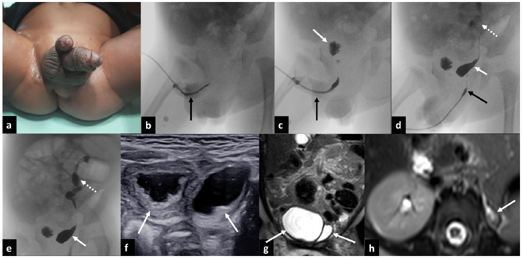
- (a) Clinical photograph showing diphallia. (b-c) Retrograde urethrogram of the upper phallus reveals opacification of a distinct urethra (black arrows) and communication with one of the two separate urinary bladders (white arrow in c). (d-e) Retrograde urethrogram of the lower phallus shows opacification of the second distinct urethra (black arrow in d) and its communication with the second urinary bladder (white arrows in d and e). There is also contrast opacification of the dilated and tortuous left ureter and left pelvicalyceal system, indicative of vesicoureteral reflux (dotted arrows in d and e). (f) Gray-scale ultrasound showing duplicated urinary bladders (white arrows). (g-h) Magnetic resonance imaging confirming duplicated urinary bladders (white arrows in g) and a small left kidney (white arrow in h).
Diphallia is a rare anomaly, with an incidence of 1 in 5–6 million live births. It is an embryological malformation with complex etiologies, often related to gene alterations.1 Diphallia can be associated with various abnormalities, including genitourinary, gastrointestinal, spinal, cardiovascular, and musculoskeletal abnormalities. Genitourinary anomalies may include duplicated urethra and urinary bladder, ectopia vesicae, VUR, single functioning kidney, and pelvic kidney.2
Conflicts of interest
There are no conflicts of interest.
References
- Diphallia with associated anomalies: A case report and literature review. Case Rep Urol. 2013;2013:192960.
- [CrossRef] [PubMed] [PubMed Central] [Google Scholar]
- Diphallia: Literature review and proposed surgical classification system. ANZ J Surg. 2022;92:2053-65.
- [CrossRef] [PubMed] [PubMed Central] [Google Scholar]


