Translate this page into:
Hydatid cyst of urinary bladder
Address for correspondence: Mr. F. A. Ganie, Department of Cardiovascular and Thoracic Surgery, SKIMS, Soura - 190 011, Kashmir, India. E-mail: farooq.ganie@ymail.com
This is an open-access article distributed under the terms of the Creative Commons Attribution-Noncommercial-Share Alike 3.0 Unported, which permits unrestricted use, distribution, and reproduction in any medium, provided the original work is properly cited.
This article was originally published by Medknow Publications & Media Pvt Ltd and was migrated to Scientific Scholar after the change of Publisher.
Sir,
Hydatid cyst of liver and lungs are common, but hydatid cysts have been seen on rare occasions at unusual sites, spleen, kidney, muscle, brain, spine, breast, thyroid, peritoneal cavity, and retroperitoneum.[1] Hydatid cyst of urinary bladder (UB) is very rare.[23]
A 48-year-old-male, known case of carcinoma larynx (post-laryngectomy) presented with a history of lower abdominal pain, urgency and feeling of incomplete urination for last 2 years. There was no history of fever, hematuria or pyuria. However, he had a history of hydatid cyst liver for which he had undergone surgery 10 years back.
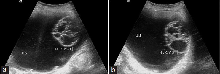
On examination, routine blood tests and chest X-ray were normal. Ultrasound abdomen showed multi-loculated cystic lesion measuring 5.2 cm × 4.8 cm arising from the left antero-lateral wall of UB [Figure 1a and b]. The lesion had typical “cyst within cyst appearance,” also called “spoke in wheel” appearance. Ultrasound (USG) guided fine needle aspiratioon (FNA) was carried out, which showed clear fluid, but microscopy was inconclusive. Based on the typicall USG findings, a provisional diagnosis of hydatid cyst of UB was made. Surgical excision of the cyst was carried out [Figure 2]. Post-operative period was uneventful. Histopathology confirmed the diagnosis.

- (a, b) 48-year-old man with hydatid cyst urinary bladder. USG shows multilocular cystic lesion “with cyst within cyst appearance” (UB: Urinarybladder, H: Hydatid cyst)

- Excisied hydatid cyst
The pathogenesis of bladder hydatid cyst is explained by hematogenous spread with development of cyst in the wall of UB. The bladder hydatid cyst clinically remains silent for long periods.[4] However, patient may present with acute retention of urine, lower abdomen pain urgency or hydaturia. In our case, patient had lower abdominal pain, urgency, and sense of incomplete urination.
USG is effective in the diagnosis of hydatid cysts. USG findings in hydatid cysts are of five types; (1) simple cyst with no internal architecture, (2) cyst with echogenic sand, (3) cyst with floating membranes (detached endocyst) also called as “water Lily sign,” (4) multi-locular cyst, which gives “cyst within cyst appearance” and (5) calcified cyst. Aspiration of fluid under USG guidance is safe, simple, and effective in reaching out provisional diagnosis.[5] Serological test are helpful in difficult cases.
The treatment of hydatid cyst is primarily surgical. However, both pre- and post-operative medical treatment facilitates the operation and decrease the recurrence.
References
- Surgical Diseases in the Tropics. Bombay, Calcutta, Madras: Macmillan India Ltd; 1982. p. :151-69.
- [Google Scholar]
- Hydatid cyst of the kidney. A report of 147 controlled cases. Eur Urol. 2000;38:461-7.
- [Google Scholar]
- Unintended percutaneous aspiration of pulmonary echinococcal cysts. AJR Am J Roentgenol. 1984;143:123-6.
- [Google Scholar]


