Translate this page into:
Kidney Dimensions and its Correlation with Anthropometric Parameters in Healthy North Indian Adults
Corresponding author: Sanjay D’Cruz, Department of General Medicine, Government Medical College and Hospital, Chandigarh, India. Email: sanjaydcruz@gmail.com
-
Received: ,
Accepted: ,
How to cite this article: Bhardwaj S, Singh A, Kaur R, D`Cruz S. Kidney Dimensions and its Correlation with Anthropometric Parameters in Healthy North Indian Adults. Indian J Nephrol. 2024;34:636-42. doi: 10.25259/ijn_12_24
Abstract
Background:
Knowledge of kidney size is important in the assessment of kidney function. Changes in kidney size can occur in various kidney diseases due to different causes, hence knowledge of normal kidney dimensions in a population is crucial for diagnosis, follow-up and prognostication. While data from other parts of the world does not apply to the Indian population due to differences in ethnicity, diet and body sizes, and there is also a lack of standardized data on normal kidney sizes in healthy Indian adults.
Materials and Methods:
Kidney dimensions from 600 healthy adult volunteers ranging between 20 and 70 years of age were measured with sonography by a single radiologist. Differences in dimensions between men and women, and right and left kidney were analyzed. Finally, kidney sizes were correlated with anthropometric variables such as weight, age, body surface area (BSA), height and body mass index. Estimated glomerular filtration rate (eGFR) was correlated with kidney length and renal parenchymal volume (RPV).
Results:
The mean kidney length of the whole cohort, irrespective of gender was found to be 9.6 ± 0.7 cm on the right and 9.9 ± 0.7 cm on the left. Mean kidney length in males was significantly more as compared to females on both sides. Both the kidney length and RPV were significantly associated with BSA, weight and height (in that order) in females, whereas in males, kidney length and RPV best correlated with height, BSA and weight (in that order). In both sexes, there was a significant negative correlation between age and kidney length, RPV. eGFR had a significant positive correlation with kidney length and RPV in the cohort.
Conclusion:
Normal sonographic mean kidney length was 9.6±0.7 cm and 9.9±0.7 cm on the right and left sides respectively in healthy North Indian population, with the left kidney being larger than the right in all dimensions (length, width, thickness and RPV). Kidney sizes in males were found to be larger than females. Correlation with anthropometric parameters in our study, emphasizes the need to give due consideration to normal variations in kidney sizes with age, gender, height, weight and BSA to differentiate between a normal and a pathologically small or large kidney.
Keywords
Kidney
Length
Ultrasound
Renal parenchymal volume
Kidney volume
Healthy adults
Introduction
Kidney size and function reflect kidney health.1 Changes in renal dimensions on ultrasound are important in determining kidney disease, as kidney sizes are considerably influenced by structural urinary tract diseases, systemic diseases, congenital anomalies, renovascular diseases1 and malignancy.
The assessment of kidney size is the starting point of evaluation of renal diseases for both diagnosis and prognostication. Ultrasound (USG) is a basic modality available at most centers, is non-invasive for measuring location and size of the kidneys and detecting any focal lesions. However, sonography has been shown to underestimate kidney volume as compared to measurements by magnetic resonance imaging and computed tomography.2 Nevertheless, because of its low cost, safety and ubiquitous availability, USG is widely accepted and considered as the initial modality of choice, especially in situations where repeat examinations are mandated.3
Kidney size measurement entails measuring the length, total volume, cortical volume and thickness. Of clinical relevance are kidney length and volume which serve as surrogates for renal functional reserve and are used frequently as the basis for making clinical decisions.4
In subjects with normal kidney function, the most frequently measured parameter is the longitudinal dimension (length) of kidney. However, studies have shown that renal parenchymal volume (RPV) is a more accurate USG parameter in end-stage renal failure.5 Kidney volume is a labor-intensive and time-consuming process and is correlated with subject’s height (ht), weight (wt) and total body surface area (BSA), but it is subject to high interobserver variations due to three parameters being involved. More recent literature suggests that kidney cortical thickness measured on ultrasound is a better indicator of kidney function in chronic kidney disease than length, and is more closely related to e-glomerular filtration rate (eGFR).6
Age-related nomograms are most commonly used to interpret normal kidney length. As kidney size abnormalities occur as a result of various kidney diseases, having established USG nomograms becomes valuable when dealing with such patients in a local setting. Data on normal kidney sizes are available from the Western population.7 However, data from the Indian subcontinent is sparse regarding kidney sizes and its correlation with other somatic parameters in the healthy adult population.8
The data available in the Western literature cannot be extrapolated to the Indian population since the kidney sizes may differ due to differences in ethnicity, fetal environment, body size, varying lifestyle factors such as high salt diet and a lower measured GFR.7,9-11 Population-based studies are needed to establish the normal values for Indian subjects. In this study, we aimed to establish standardized data for normal kidney dimensions on sonography in the Indian population and correlate it with age, sex, height, weight, body mass index, BSA and eGFR. We also aimed to formulate a nomogram of the normal kidney size in the North Indian population for further comparison.
Materials and Methods
After obtaining clearance from the institutional ethics committee, an age and sex stratified random sample of 600 men and women ranging between 20 and 70 years were taken over a period of one-and-a-half year, with the age strata in years being 20–35, 36–50 and >50. All eligible individuals were chosen from the North Indian population, which included healthy volunteers accompanying patients, nurses and students. Healthy volunteers were selected on the basis of normal blood pressure, random blood sugar, urinalysis and serum creatinine.
Subjects with known renal disease and symptomatic renal calculus disease, and those with eGFR≤60 mL/min/1.73 m2, known kidney disease or any structural kidney condition found incidentally on ultrasound like hydronephrosis, horseshoe kidney and duplicate ureters and with diabetes mellitus and hypertension were excluded.
All the respondents provided informed written consent. Their demographic details, height and weight, and relevant medical histories were noted.
All ultrasound examinations were performed on a Phillips IU 20 or Toshiba Nemio 30 system using a 3.5 MHz convex array transducer by a single experienced radiologist (all participants were asked to empty the bladder just before the examination).
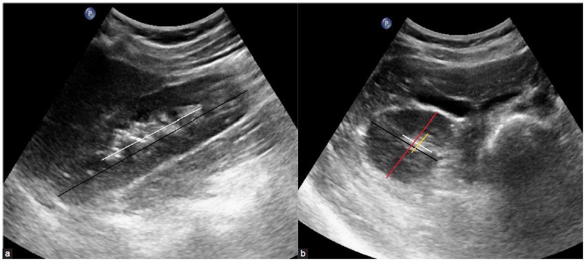
Maximum longitudinal pole-to-pole length was taken for each kidney in the sagittal plane, either in the supine position or by slightly elevating the site of examination after re-positioning the probe so that the section represented the maximum longitudinal dimension of the kidney. The length of the central echogenic area was also taken in the same plane. The width and thickness of the kidney and central echogenic area were taken in a plane orthogonal to the longitudinal section close to the renal hilum and excluding the renal pelvis in measurements. Kidney width was measured as the maximum distance between the medial and lateral borders of the kidney. In the same plane, renal thickness was also measured as the distance between ventral and dorsal surfaces of the kidney. Width and thickness of the central echogenic area were also measured in the same plane [Figure 1].

- (a) Measurement of renal length (black line) and central echogenic area (white line) in the longitudinal section of the kidney. (b) Width (black line) and thickness (red line) of the kidney and central echogenic area (white and yellow line, respectively) in the axial section of the kidney.
Mean of three readings was taken for each measurement. Volume of the kidneys was measured by the appropriate formula.
The renal volume and volume of the central echogenic area was calculated by the formula: Volume = 0.5233 × (Length × Width × Thickness)
RPV = (Renal volume − Central echogenic volume)
Total BSA, body mass index and eGFR were calculated as follows:
BSA = Weight0.425 × Height0.725 × 71.84.
Body mass index (BMI) = Weight (kg)/Height (m2)
eGFR was calculated using CKD-EPI Creatinine 2021 equation- eGFR
Correlation of renal length and RPV with age, gender, height, weight, BMI, BSA and eGFR was determined.
Data was entered in a Microsoft Excel sheet and statistically analyzed using Statistical Package for Social Sciences (SPSS) Version-23. Comparative analyses were done by means of appropriate statistical tools like student’s “t” test, Chi square test, Mann–Whitney test, Analysis of variance (ANOVA) and Pearson correlation coefficient (r) as applicable. A p-value < 0.05 was regarded as statistically significant.
Results
Of the 600 subjects enrolled for the study, 89 were excluded for the following reasons: hypertensive (20), diabetic (25), proteinuria (24) and renal calculi (10), hydronephrosis (6) and polycystic kidney (4).
Ultrasound was thus performed on 511 healthy individuals who met the inclusion criteria, which included 309 males and 202 females with no known renal disease or dysfunction.
The anthropometric measurements of the study population are summarized in Table 1.
| Variable | Gender | Mean ± (SD) | SEM | p-value | |
|---|---|---|---|---|---|
| Age (yrs) | F | 38.71 | 12.56 | .884 | 0.802 |
| M | 38.99 | 12.13 | .690 | ||
| Weight (kg) | F | 58.15 | 12.73 | .896 | 0.024 |
| M | 60.61 | 11.42 | .650 | ||
| Height (cm) | F | 159.18 | 6.07 | .427 | <0.001 |
| M | 166.56 | 6.73 | .383 | ||
| Body mass index (kg/m2) | F | 22.94 | 4.92 | .346 | 0.003 |
| M | 21.81 | 3.69 | .210 | ||
| Body surface area | F | 1.59 | 0.16 | .012 | <0.001 |
| M | 1.67 | 0.16 | .009 | ||
| Urea (mg/dl) | F | 23.75 | 7.68 | .541 | 0.001 |
| M | 25.99 | 7.54 | .429 | ||
| Creatinine (mg/dl) | F | .65 | 0.20 | .014 | <0.001 |
| M | .75 | 0.23 | .013 | ||
| eGFR (ml/min/1.73m2) | F | 123.05 | 44.34 | ||
| M | 134.4 | 44 | |||
SEM: Standard error of measurement, eGFR: Estimated glomerular filtration rate.
The mean kidney length was 9.56 ± 0.7 cm on the right and 9.95 ± 0.7 cm on the left side. The mean kidney width and thickness were 4.8 ± 0.6 cm and 4.25 ± 0.6 cm, and 4.75 ± 0.6 cm and 4.3 ± 0.6 cm on the right and left side, respectively. The estimated average RPV was 91.49 ± 24.4 cm3 on the right and 93.95 cm3 on the left side [Table 2].
| Parameter | Males | Females | 20–35 yrs (n = 243) | 36–50 yrs (n = 174) | >50 yrs (n = 94) | All (511) | |
|---|---|---|---|---|---|---|---|
|
Length Mean ±SD |
Right | 9.64 ± 0.68 |
9.51 ± 0.69 p = 0.001 |
9.67 ± 0.69 p = 0.001 |
9.61 ± 0.62 p = 0.001 |
9.34 ± 0.74 p = 0.001 |
9.60 ± 0.68 |
| Left | 10.04 ± 0.72 |
9.83 ± 0.77 p < 0.001 |
10.02 ± 0.05 p = 0.001 |
9.99 ± 0.05 P = 0.001 |
9.68 ± 0.08 p = 0.001 |
9.95 ± 0.74 | |
|
Width (cm) Mean ±SD |
Right | 4.83 ± 0.60 |
4.63 ± 0.6 p < 0.001 |
4.78 ± 0.59 p = 0.003 |
4.84 ± 0.57 p = 0.003 |
4.53 ± 0.45 p = 0.003 |
4.75 ± 0.64 |
| Left | 4.91 ± 0.60 |
4.73 ± 0.53 p < 0.001 |
4.83 ± 0.54 p = 0.001 |
4.80 ± 0.58 p = 0.001 |
4.65 ± 0.61 p = 0.001 |
4.82 ± 0.68 | |
|
Thickness (cm) Mean ± SD |
Right | 4.40 ± 0.60 |
4.00 ± 0.54 p < 0.001 |
4.27 ± 0.56 | 4.28 ± 0.63 | 4.16 ± 0.64 | 4.26 ± 0.61 |
| Left | 4.37 ± 0.58 |
4.10 ± 0.52 p < 0.001 |
4.29 ± 0.58 | 4.28 ± 0.57 | 4.25 ± 0.57 | 4.28 ± 0.57 | |
|
Length (cm) (central echogenic area) Mean ± SD |
Right | 6.35 ± 0.71 | 6.24 ± 0.72 | ||||
| Left | 6.59 ± 0.68 | 6.47 ± 0.72 | |||||
|
Width (cm) (central echogenic area) Mean ± SD |
Right | 2.20 ± 0.42 | 2.10 ± 0.44 | ||||
| Left | 2.10 ± 0.40 | 2.10 ± 0.41 | |||||
|
Thickness (cm) (central echogenic area) Mean ±SD |
Right | 1.90 ± 0.43 | 1.70 ± 0.41 | ||||
| Left | 1.90 ± 0.41 | 1.70 ± 0.41 | |||||
|
Volume Total (cm3) Mean ± SD |
Right | 110 ± 28 | 95 ± 23.1 | ||||
| Left | 112.3 ± 27 | 99.3 ± 24 | |||||
|
Volume central echogenic area (cm3) Mean ± SD |
Right | 13.7 ± 5.6 | 11.7 ± 5.2 | ||||
| Left | 13.9 ± 4.9 | 12.2 ± 4.5 | |||||
|
Renal parenchymal volume (cm3) Mean ± SD |
Right | 96.8 ± 25.3 |
83.2 ± 20.4 p < 0.001 |
92.44 ± 23.13 p = 0.007 |
93.83 ± 24.87 p = 0.007 |
84.39 ± 25.56 p = 0.007 |
91.44 ± 24.4 |
| Left | 98.4 ± 24.2 |
87.13 ± 21.2 p < 0.001 |
95.33 ± 24.11 p = 0.001 |
95.98 ± 23.62 p = 0.004 |
86.58 ± 21.59 p = 0.004 |
93.95 ± 23.72 | |
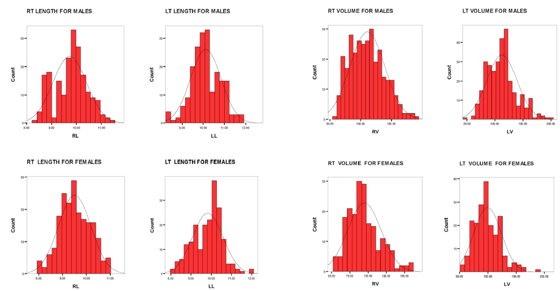
Mean kidney lengths in males were significantly higher than in females on both sides. It was 9.6 ± 0.7 (males) and 9.5 ± 0.7 (females) respectively on the right side and 10.0 ± 0.72 and 9.8 ± 0.8 respectively on the left side. The nomograms for right and left kidney lengths in both genders are given in Figure 2. Mean renal widths were also significantly more in males than females viz: 4.9 ± 0.6 cm (males) and 4.7 ± 0.53 cm (females) on right side and 4.8 ± 0.56 (males) and 4.6 ± 0.6 cm (females) on left side. Similarly, there was a statistically significant difference in mean renal thickness in males and females i.e. 4.4 ± 0.6 cm (males) and 4.0 ± 0.54 (females) on right side and was 4.4 ± 0.6 cm (males) and 4.1 ± 0.52 cm (females) on left side respectively [Table 2].

- Nomograms for right and left kidney lengths and right and left RPV in males (upper panel) and females (lower panel). RT: right, LT: left, RL: Right length, LL: left length, RV: right volume and LV: left volume. RPV: renal parenchymal volume.
Right RPV was higher in males, 96.8 ± 25.3 cm3 and 83.2 ± 20.4 cm3 in males and females, respectively. Left RPV was also higher in males, 98.4 ± 24.2 cm3 and 87.1 ± 21.2 cm3 in males and females, respectively. The nomograms for right and left RPV in both genders are given in Figure 2.
ANOVA revealed the differences in mean renal length, renal width and RPV in different age groups 20–35, 36–50 and >50 years to be statistically significant on bothsides [Table 2].
In females, renal length best correlated with BSA with a significant (r) of 0.245 and 0.166 on the right and left sides respectively, followed by weight with a significant (r) of 0.224 and 0.156 on the right and left side respectively, followed by height with a significant (r) of 0.205 and 0.145 on the right and left side respectively. Renal length showed a negative correlation with age, –0.053 on the right and –0.125 on the left side but was not statistically significant. There was no correlation of renal length with BMI.
In females, RPV best correlated with BSA with a significant (r) of 0.242 and 0.248 on right and left sides, respectively, followed by weight with a significant (r) of 0.231 and 0.248 on right and left sides, respectively, followed by height having a significant (r) of 0.200 and 0.242 on the right and left sides, respectively. RPV showed a negative correlation with age, having a coefficient of –0.048 on the right and –0.013 on the left side but were not statistically significant. RPV showed no correlation with BMI.
In males, renal length best correlated with height [significant (r) of 0.158 and 0.125 on the right and left sides, respectively], followed by BSA [significant (r) of 0.139 and 0.113 on the right and left sides, respectively]followed by weight [significant (r) of 0.113 and 0.111 on the right and left sides, respectively]. Renal length showed a significant negative correlation with age, with a (r) of –0.181 and –0.188 on the right and left sides, respectively. Renal length in males did not correlate with BMI.
In males, RPV best correlated with height with a significant (r) of 0.166 and 0.116 on the right and left sides, respectively, followed by BSA with a significant (r) of 0.135 and 0.178 on the right and left sides, respectively, followed by weight having a significant (r) of 0.117 and 0.190 on the right and left sides, respectively. RPV showed a significant negative correlation with age, with a coefficient of –0.142 and –0.177 on the right and left sides, respectively. RPV in males showed no correlation with BMI.
Finally, eGFR positively correlated with mean RPV (r = 0.093, p-value 0.035).
Discussion
We aimed to establish reference ranges regarding normal kidney sizes measured by ultrasound in the healthy North Indian adult population and correlate it with somatic and physiological parameters such as height, age, weight, BSA, BMI and eGFR in males and females. Kidney dimensions are important for studying renal function and its disorders. They are valuable for making a primary diagnosis and disease follow-up. Although some papers look at kidney sizes in the Indian population, our study is the largest in terms of sample size. As compared to other studies, we have excluded patients with diabetes, hypertension, asymptomatic urinary abnormalities and eGFR < 60 mL/min, which makes our data more robust and representative of normal population.
To our knowledge, our study is the largest to have all measurements done by a single radiologist, eliminating interobserver variability. Also, we eliminated intra-observer variability by taking a mean of 3 readings for each measurement. Our work focused not only on linear renal dimensions, but also on three-dimensional volumetric data. Though our study was based in a tertiary care hospital, the healthy participants represented a fair cross section of the general population. Finally, we also correlated eGFR with mean kidney dimensions.
Our efforts stem from the understanding that kidney sizes are different amongst different populations across the globe. Papers from different parts of the world have reported larger normal kidney sizes compared to our findings and other studies from the Southeast Asian population. The mean kidney size of 9.89 + 0.9 cm in 100 healthy kidney donors assessed on CT in a study from North India is similar to our results (9.90 cm + 0.7).12 Differences in kidney dimensions in different populations reflects differences in ethnicity, dietary habits, body size and habitus, thereby emphasizing the need to have a normal nomogram for our set of patients. Organ size unquestionably relates to body size and our findings reflect the relatively small body size of the average Indian.13 Larger kidney sizes have been found in studies done in the Nigerian, Caucasian, Mexican and Iranian populations.3,10,14-16 Table 3 summarizes the kidney lengths from other studies including our observations.17-19
| Country | Total Number of Subjects | Renal Lengths (cm) | |||
|---|---|---|---|---|---|
| Males | Females | All | |||
| Index Study | 511 | Right | 9.64 | 9.51 | 9.6 |
| Left | 10.04 | 9.83 | 9.9 | ||
| Iran18 | 400 | Right | 11.0 | 10.7 | 10.9 |
| Left | 11.3 | 10.9 | 11.1 | ||
| Malaysia10 | 205 | Right | 10.2 | 9.8 | - |
| Left | 10.5 | 10.0 | - | ||
| Mexico14 | 153 | Right | 10.57 | 10.29 | 10.43 |
| Left | 10.72 | 10.46 | 10.58 | ||
| Nigeria15 | 200 | Right | - | - | 10.3 |
| Left | - | - | 10.6 | ||
| USA19 | - | Right | - | - | 10.7 |
| Left | 11.1 | ||||
| Pakistan3 | 194 | Right | 10.6 | 10.3 | 10.45 |
| Left | 10.6 | 10.3 | 10.45 | ||
We noted that the left kidney was significantly larger than the right in length, width, thickness and RPV. Possible explanations include more space available for growth to the left kidney due to smaller size of the spleen compared to the liver, and a more straight, short course of the left renal artery contributing more blood flow compared to the right.20 The same has been reported in multiple papers by other researchers.7,10,21-23 We also found larger kidney sizes in males compared to females with respect to the RPV, length, width and thickness, however, these differences were unadjusted for differences in height, weight and BSA. Most of the studies report mean kidney length to be larger in males as compared to females. They probably result from the larger anthropometric dimensions in males as compared to females. However, some studies report no difference between the two sexes.24
Kidney reaches its mature size at age 20–29 years and remains relatively unchanged until the sixth decade of life. Studies show that aging leads to progressive decrease in kidney size, after middle age3,21,23,25 at a rate of 0.5 cm per decade, especially due to a reduction of blood flow by 1% per year after the third decade.26 It is well established that by 70 years, as much as 30–50% of the cortical glomeruli atrophy; manifested by a progressive loss in renal mass.27 We saw a weak negative correlation between renal length, RPV and age, which was significant in males but non-significant in females. A smaller number of participants over the age of 70 and a broader age stratum could explain these findings.
We also correlated kidney dimensions with various anthropometric measures. Renal length and RPV were positively correlated with height, BSA and weight in both sexes, however the order varied between the two sexes. Some papers have reported renal volume to be the most accurate when correlated with body weight; normal values of total renal volume per kg of body weight were 4.3–8.0 mL/kg.24 According to our data, the most significant factors associated with kidney size were sex, weight, height and BSA. While the strength of correlation of individual sonographic measures with different anthropometric measures might differ in males and females, and some parameters might have a bigger impact than others, we believe it’s the combination of all these anthropometric measurements which ultimately determines kidney size in a healthy individual.
Our study has certain limitations. We had few participants over 70, possibly failing to detect a statistically significant correlation between kidney length, RPV and age. The participants were limited to Northern India, which might not be a holistic representation of the entire Indian population. A bigger nationwide study with an even larger sample size could find significant correlations which our study failed to detect. This study cannot serve as a base for a nomogram for the entire Indian population, for which a larger number of healthy individuals in each group and across various Indian ethnicities and regions will be required. Finally, age could have been a confounder in correlations between eGFR and kidney dimensions, and while statistical significance was achieved, the results offer doubtful clinical significance.
This study provided data for normal sonographic renal dimensions in a North Indian population with a mean kidney length of the whole cohort.
Conclusions about renal sizes need to be made concerning nomograms and should not be extrapolated from data from other populations. Irrespective of gender, the left kidney was found to be larger than the right in all kidney dimensions (length, width, thickness and RPV). Kidney dimensions in males were found to be larger than females. The paper also provides data regarding the positive correlation between eGFR with renal dimensions including kidney length and RPV in healthy adults.
While interpreting a kidney ultrasound, variations of renal size with age, gender, height, weight and BSA should be considered by the clinician to differentiate between a normal and a pathologically small or large kidney.
Conflicts of interest
There are no conflicts of interest.
References
- SMART Study Group. Influence of atherosclerosis on age-related changes in renal size and function. Eur J Clin Invest. 2003;33:34-40.
- [CrossRef] [PubMed] [Google Scholar]
- In vitro measurement of kidney size: comparison of ultrasonography and MRI. Ultrasound Med Biol. 1998;24:683-8.
- [CrossRef] [PubMed] [Google Scholar]
- Ultrasonographic renal size in individuals without known renal disease. J Pak Med Assoc. 2000;50:12-6.
- [PubMed] [Google Scholar]
- Value of routine renal and abdominal ultrasonography in patients undergoing prostatectomy. Int Urol Nephrol. 2006;38:153-6.
- [CrossRef] [PubMed] [Google Scholar]
- Confronto tra biometria ecografica e funzionale renale in pazienti sani e in pazienti con IRC [Comparison of renal ultrasonographic and functional biometry in healthy patients and in patients with chronic renal failure] Arch Ital Urol Androl. 2002;74:206-9. Italian
- [PubMed] [Google Scholar]
- Renal cortical thickness measured at ultrasound: is it better than renal length as an indicator of renal function in chronic kidney disease? AJR Am J Roentgenol. 2010;195:W146-9.
- [CrossRef] [PubMed] [Google Scholar]
- Kidney dimensions at sonography: Correlation with age, sex, and habitus in 665 adult volunteers. AJR Am J Roentgenol. 1993;160:83-6.
- [CrossRef] [PubMed] [Google Scholar]
- Ultrasonographic assessment of renal size and its correlation with body mass index in adults without known renal disease. J Ayub Med Coll Abbottabad. 2011;23:64-8.
- [PubMed] [Google Scholar]
- Morphométrie du rein. Etude appliguée à l’urologie et à l’imagerie [Morphometry of the kidney. Applied study in urology and imaging] J Urol (Paris). 1989;95:77-80. French
- [PubMed] [Google Scholar]
- Renal size in healthy malaysian adults by ultrasonography. Med J Malaysia. 1989;44:45-51.
- [PubMed] [Google Scholar]
- A study of normal renal dimensions at ultrasonography and their influencing factors in an Indian population. Cureus. 2023;15:e40748.
- [CrossRef] [PubMed] [PubMed Central] [Google Scholar]
- Estimation of renal length in adult north indian population: A ct study. Int J Anat Res. 2016;4:1837-42.
- [Google Scholar]
- Weight and measurements of kidneys in northwest Indian adults. Am J Hum Biol. 2001;13:726-32.
- [CrossRef] [PubMed] [Google Scholar]
- Longitud renal por ultrasonografía en población mexicana adulta [Renal length measured by ultrasound in adult mexican population] Nefrologia. 2009;29:30-4. Spanish
- [Google Scholar]
- Normal sonographic renal length in adult southeast Nigerians. Afr J Med Med Sci. 2005;34:129-31.
- [PubMed] [Google Scholar]
- Roentgenologic estimation of kidney size in adult Nigerians. Trop Geogr Med. 1982;34:177-81.
- [PubMed] [Google Scholar]
- Need for a nomogram of renal sizes in the Indian population- findings from a single centre sonographic study. Indian J Med Res. 2014;139:686-93.
- [PubMed] [PubMed Central] [Google Scholar]
- Sonographic measurement of absolute and relative renal length in healthy Isfahani adults. J Res Med Sci. 2004;9:54-7.
- [Google Scholar]
- Ultrasound assessment of normal renal dimensions. J Ultrasound Med.. 1982;1:49-52.
- [CrossRef] [PubMed] [Google Scholar]
- Kidney dimensions at sonography: correlation with age, sex, and habitus in 665 adult volunteers. AJR Am J Roentgenol.. 1993;160:83-6.
- [CrossRef] [PubMed] [Google Scholar]
- Normal renal dimensions in a specific population. Int Braz J Urol. 2002;28:510-5.
- [PubMed] [Google Scholar]
- A sonographic study of kidney dimensions in a sample of healthy Jamaicans. West Indian Med J. 2000;49:154-7.
- [PubMed] [Google Scholar]
- Sonographic measurement of absolute and relative renal length in adults. J Clin Ultrasound. 1998;26:185-9.
- [CrossRef] [PubMed] [Google Scholar]
- Determination of renal volume by ultrasound scanning. J Clin Ultrasound. 1978;6:160-4.
- [CrossRef] [PubMed] [Google Scholar]
- Sonographic measurement of kidney size in geriatric patients. J Clin Ultrasound. 2003;31:315-8.
- [CrossRef] [PubMed] [Google Scholar]
- Changes in sizes and distensibility of the aging kidney. Br J Radiol. 1981;54:488-91.
- [CrossRef] [PubMed] [Google Scholar]
- Cell senescence and its implications for nephrology. J Am Soc Nephrol. 2001;12:385-93.
- [CrossRef] [PubMed] [Google Scholar]


