Translate this page into:
Pancake Kidney: A Rare Case of Renal Ectopia
Corresponding author: Abhishek Pratap Singh, Department of Nephrology, SMS Medical College, Jaipur, India. E-mail: abhishekpratapsingh85@gmail.com
-
Received: ,
Accepted: ,
How to cite this article: Singh AP, Beniwal P, Malhotra V. Pancake Kidney: A Rare Case of Renal Ectopia. Indian J Nephrol. 2025;35:111-2. doi: 10.25259/IJN_401_2024
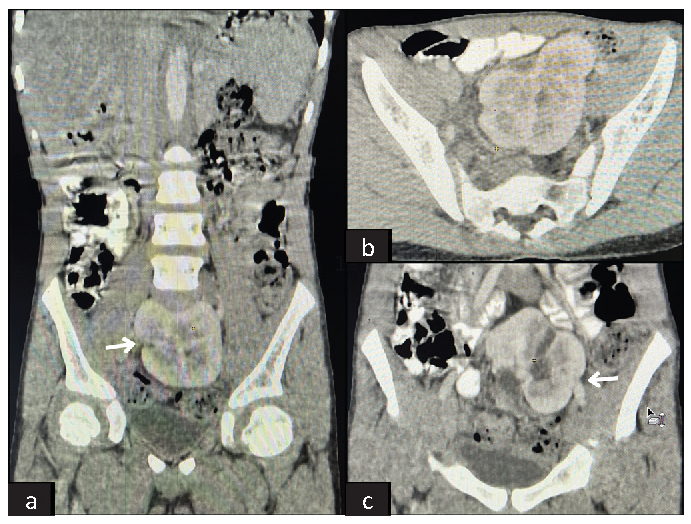
An 11-year-old male presented with a 1-month history of lower abdominal pain, accompanied by fever and burning micturition for the past 7 days. Abdominal examination was unremarkable, with no tenderness at the bilateral renal angles. Urine microscopy revealed pyuria, though kidney function tests and X-rays of the kidney, ureter, and bladder (KUB) were normal. Ultrasonography of the KUB region identified an abnormally located kidney in the pelvis. Computed tomography (CT) urography further revealed that both kidneys were fused in the pelvic cavity at the midline, with a single mega ureter on the left side, suggestive of a pancake kidney [Figure 1]. He was prescribed a course of antibiotics based on urine culture and sensitivity test results, which successfully relieved his symptoms.

- (a-b) Computed Tomography (CT) urography images showing (white arrow in a) the complete medial fusion of the renal parenchyma, located ectopically in the pelvic cavity at the midline. (c) CT urography image (white arrow) showing a single megaureter originating from the left side of the fused renal mass.
Pancake kidney is an exceptionally rare form of fused renal ectopia. Looney and Dodd were the first to define and describe this condition.1 It is characterized by a renal mass in the pelvis resulting from the complete medial fusion of the renal parenchyma without an intervening septum. Typically, each lobe has a separate pelvicalyceal system, but in our case, the kidneys had a single megaureter entering the bladder at the left vesicoureteral junction.
This anomaly can predispose to recurrent urinary tract infections and stone formation due to probable rotation anomalies of the collecting system and short ureters, which are prone to stasis and obstruction. Congenital renal malformations are often incidentally detected and asymptomatic; in such cases, a conservative approach with long-term follow-up is recommended.2
Declaration of patient consent
The authors certify that they have obtained all appropriate patient consent.
Conflicts of interest
There are no conflicts of interest.
References
- An ectopic (pelvic) completely fused (cake) kidney associated with various anomalies of the abdominal viscera. Ann Surg. 1926;84:522-4.
- [CrossRef] [PubMed] [PubMed Central] [Google Scholar]
- Pancake kidney found inside abdominal cavity: Rare case with literature review. Urol Case Rep. 2017;13:123-5.
- [CrossRef] [PubMed] [PubMed Central] [Google Scholar]


