Translate this page into:
Th1, Th2 and Treg/T17 cytokines in two types of proliferative glomerulonephritis
Address for correspondence: Dr. M. Stangou, Department of Nephrology, Aristotle University, Hippokration Hospital, Thessaloniki, Greece. E-mail: mstangou@auth.gr
This is an open access article distributed under the terms of the Creative Commons Attribution-NonCommercial-ShareAlike 3.0 License, which allows others to remix, tweak, and build upon the work non-commercially, as long as the author is credited and the new creations are licensed under the identical terms.
This article was originally published by Medknow Publications & Media Pvt Ltd and was migrated to Scientific Scholar after the change of Publisher.
Abstract
IgA nephropathy (IgAN) and focal segmental necrotizing glomerulonephritis (FSNGN) are characterized by proliferation of native glomerular cells and infiltration by inflammatory cells. Several cytokines act as mediators of kidney damage in both diseases. The aim of the present study was to investigate the role of Th1, Th2 and Treg/T17 cytokines in these types of proliferative glomerulonephritis. Simultaneous measurement of Th1 interleukin (IL-2, IL-12, tumor necrosis factor-alpha [TNF-α], interferon-gamma [INF-γ]), Th2 (IL-4, IL-5, IL-6, IL-10, IL-13), Treg/T17 transforming growth factor-beta 1 (TGF-β1, granulocyte-macrophage colony-stimulating factor [GM-CSF], IL-17) cytokines and C-C chemokines Monocyte chemoattractant protein-1 (MCP-1, macrophage inflammatory protein-1 [MIP-1] β) was performed in first-morning urine samples, at the day of renal biopsy, using a multiplex cytokine assay. Cytokine concentrations were correlated with histological findings and renal function outcome. Urinary excretion of Th1, Th2 and Treg/Th17 cytokines were significantly higher in FSNGN compared to IgAN patients. In IgAN patients (n = 50, M/F: 36/14, M age: 40.7 [17–67] years), Th1, Th2 and T17 cytokines correlated significantly with the presence of endocapillary proliferation, while in FSNGN patients (n = 40, M/F: 24/16, M age: 56.5 [25–80] years), MCP-1 and TGF-β1 had a positive correlation with severe extracapillary proliferation (P = 0.001 and P = 0.002, respectively). Urinary IL-17 was the only independent parameter associated with endocapillary proliferation in IgAN and with MCP-1 urinary excretion in FSNGN. Response to treatment was mainly predicted by IL-6 in IgAN, and by Th2 (IL-4, IL-6), Treg (GM-CSF) cytokines and MIP-1 β in FSNGN. Th1, Th2 and T17 cytokines were directly implicated in renal pathology in IgAN and possibly through MCP-1 production in FSNGN. IL-17 and IL-6 seem to have a central role in inflammation and progression of kidney injury.
Keywords
Focal segmental necrotizing glomerulonephritis
IgA nephropathy
inflammation
proliferation
Th1/Th2/Treg/T17 cytokines
Introduction
IgA nephropathy (IgAN) and focal segmental necrotizing glomerulonephritis (FSNGN) are both proliferative glomerular diseases, which share some common histopathological characteristics such as glomerular hypercellularity and macrophage and T-cell infiltration.[12] The two diseases also have some substantial differences, mainly the mesangial immune complex deposition of IgA, C3 and less frequently IgG antibodies in IgAN and also, the type of glomerular hypercellularity, mesangial and endocapillary in IgAN, while extracapillary in FSNGN, which mainly involves infiltrating inflammatory cells such as macrophages.[34]
Increased urinary cytokine excretion has been found in both types of glomerulonephritis, and previous studies have described their possible participation in disease pathogenesis.[56] Monocyte chemoattractant protein-1 (MCP-1), the pro-inflammatory chemokine, main chemoattractant molecule for macrophages, has been described in both diseases as having a main role in the development of the disease.[7] However, data regarding the role of Th1, Th2 and the recently described Treg/Th17 cytokines in the initiation and progression of kidney damage, are sparse in both diseases.
In experimental models of crescentic glomerulonephritis, Th17-mediated immune reactions promote the early stages of disease, whereas Th1 cells predominate during the later stages and ultimately lead to real tissue damage.[89]
A vicious cycle of cytokine-chemokine feedback seems to take place in various models of autoimmune experimental glomerulonephritis. Th17 cells are not terminally differentiated cells, they can switch into Th1 cells and may be implicated in several inflammatory reactions such as production of production of pro-inflammatory cytokines and chemokines leading to recruitment of neutrophils and macrophages.[1011] This study aimed to investigate the roles of Th1, Th2 and Treg/T17 cytokines in the development and progression of these two types of proliferative glomerulonephritis.
Patients and Methods
Patients
A total of 90 patients with proliferative glomerulonephritis, 50 with biopsy-proven IgAN, and 40 with FSNGN were retrospectively studied. All patients were diagnosed during the period 2005-2010.
Inclusion criteria were age >18 years, biopsy-proven IgAN or FSNGN, no previous treatment with steroids, immunosuppressants and/or renin-angiotensin-aldosterone-system inhibitors (RAASi) and fish oils (FO).
Exclusion criteria were: Urine infection or septicemia at time of diagnosis, co-existent autoimmune disease (e.g., systemic lupus erythematosus, rheumatoid arthritis, Henoch Shönlein Purpura, etc.).
After their discharge from hospital, patients were followed-up regularly, on an out-patient basis, every month for the first year and every 3 months for the rest of follow-up period, totally 56 ± 43.8 months. Patients were censored if they reached end-stage renal disease (ESRD) and started on hemodialysis.
Measurement of urinary cytokines
A first-morning midstream urine sample was collected under sterile conditions from all patients, on the day of diagnosis and before any specific treatment was administered. Fifteen milliliters of urine samples were centrifuged at 1500 rpm, for 10 min and the supernatant was kept in −70°C, until assayed. Bio-Plex human cytokine assay was applied for the measurement of Th1: Interleukin (IL-2), IL-12, interferon-gamma (INF-γ), tumor necrosis factor-alpha (TNF-α), Th2: IL-4, IL-5, IL-6, IL-10, IL-13, Treg/Th17 cytokines: Transforming growth factor-beta 1 (TGF-β1), IL-17, Granulocyte-macrophage colony-stimulating factor (GM-CSF) and C-C chemokines: MCP-1 and macrophage inflammatory protein-1 (MIP-1) β. All cytokines were measured simultaneously in the same urine sample. Premixed beads coated with target capture antibodies were transferred to each well of a flat filter plate and washed twice with Bio-Plex wash buffer. After incubation with the samples and premixed detection antibodies, the beads were resuspended in Bio-Plex assay buffer and read on the Bio-Plex suspension array system. Data were analyzed using Bio-Plex Manager™ software with 5PL curve fitting. Midstream morning urine samples obtained from 10 healthy individuals were used as controls. Urinary cytokine levels were normalized for urinary creatinine (Ucr) concentration and were expressed as fg/mg Ucr.
Assessment of renal histology
Diagnosis of IgAN was based on optical microscopy and immunofluorescence and/or immunohistochemistry. Evaluation of renal pathology in IgAN was performed by the Oxford classification system, based on the presence or absence of mesangial hypercellularity, endocapillary proliferation and segmental glomerulosclerosis (M0-M1, E0-E1 S0-S1 respectively) and the degree of tubular atrophy/interstitial fibrosis, classified as T0, T1 and T2 affecting ≤25%, 26–50% and >50% respectively.[89]
Diagnosis of FSNGN was based on the presence of focal necrosis, extracapillary proliferation, and negative immunostaining. The extent of glomerular damage was estimated by the percentage of crescents in the glomeruli, and the severity of tubular atrophy/interstitial fibrosis was classified as T0, T1, T2 when ≤25%, 26–50% or >50% of interstitial area was respectively affected.
Treatment protocol
All IgAN patients were treated with RAASi and FO. An additional 12-month course of prednisolone (pred) (0.8 mg/Kg/day gradually tapered) was given to patients who had persistent proteinuria >1 g/24 day.
All patients with FSNGN received the same standard treatment protocol. Induction therapy; the first 3 months consisted of prednisolone (3 g IV followed by oral prednisolone 1 mg/kg/day, gradually tapered) and cyclophosphamide (dose adjusted for age and renal function). Plasmapheresis was performed in case of pulmonary hemorrhage or severe renal failure, requiring hemodialysis. Ten plasma exchanges of 3000 ml were performed within the first 20 days. Treatment was regularly applied just after renal biopsy was performed, except in 8/40 patients, who presented with severe life-threatening hemoptysis and had to be commenced on treatment before renal biopsy. Kidney biopsy in those patients was performed within the next 7 days. Induction therapy was followed by maintenance treatment, consisted of prednisolone and azathioprine or mycophenolate mofetil, for a total period of at least 2 years.
Definitions
Creatinine clearance (CrCl) was estimated based on 24 h urine collection. CrCl of <70 ml/min/1.73 m2 was regarded as renal function impairment; and CrCl < 10 ml/min/1.73 m2 ESRD, necessitating hemodialysis.
Response to treatment was defined as a stabilization of renal function to previous levels and reduction of proteinuria <1 g/24 h. Patients who at the end of follow-up had ≥50% increase in serum creatinine from baseline and/or protein excretion ≥1 g/24 day, were considered as nonresponders. Renal survival was defined as stabilization of serum creatinine (<50% increase from baseline).
Statistics
All values are expressed as mean ± standard deviation. Statistical analysis was performed using Statistical Package for the Social Sciences (SPSS) 17.0 for Windows. P < 0.05 was considered as statistically significant. Pearson and Spearman coefficients were used for the correlation between parametric and nonparametric variables respectively. Multivariate stepwise analysis was performed to estimate the independent parameters correlated with the outcome of renal function. Differences between groups were estimated by Mann–Whitney U-test.
Results
IgA nephropathy patients
Mean age of the patients with IgAN (n = 50, males/females 36/14) at time of presentation was 40.7 (range 17–67) years. Serum creatinine at presentation (Scr1) was 1.6 ± 0.9 mg/dl, CrCl1 was 64.3 ± 27 ml/min, and Upr1 1.5 ± 1.6 g/24 h. Forty-three patients had hypertension. All patients had microscopic hematuria while 10/50 had episodes of macroscopic hematuria [Table 1]. Nineteen patients were classified as M0 and 31 as M1, 37 as E0 and 13 as E1, 38 as S0 and 12 as S1; 32 patients were classified as T0, 10 as T1 and 8 as T2. Active lesions, such as mesangial hyperplasia and endocapillary proliferation predominated in 40 patients while chronic lesions, such as glomerulosclerosis and tubulointerstitial fibrosis, were more prominent in 10 patients. Urinary excretion of Th1, Th2 and Treg/T17 cytokines is shown in Table 2.

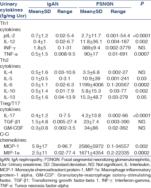
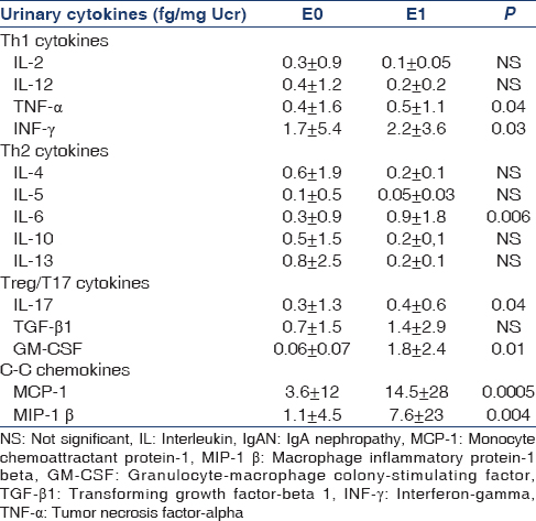
The presence of endocapillary proliferation was associated with increased urinary excretion of Th1 (INF-γ, TNF-α, P = 0.03, P = 0.04 respectively), Th2 (IL-6, P = 0.006), Th17 (IL-17, P = 0.04) and pro-inflammatory chemokines (MCP-1, MIP-1 β, P = 0.0005, P = 0.004, respectively) [Table 3]. In multiple regression analysis, IL-17 was the only independent factor correlated with the presence of endocapillary proliferation (r = 0.6, P = 0.001). The presence of mesangial hyperplasia and the degree of glomerulosclerosis and that of tubular atrophy showed no correlation with urinary cytokine levels. CrCl1 showed a significant positive correlation with the degree of proteinuria (Upr1) (r = 0.4, P = 0.02), and urinary levels of IL-2 and MCP-1 (r = 0.3, P = 0.03 and r = 0.3, P = 0.03 respectively).
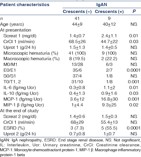
Nine IgAN patients (18%) had crescents affecting 5–30% of glomeruli on renal biopsy. Patients with crescents presented with more severe pathology and increased urinary excretion of IL-6, IL-10, MCP-1 and MIP-1 β; they had advanced renal failure at presentation and worse outcome of renal function. At the end of the follow-up, 5/9 (55.5%) of patients with crescents progressed to ESRD compared to only 3/41 (7.3%) of patients without crescents [Table 4].
At the end of the follow-up period (68 [12–155] months), 8/50 (16%) patients reached ESRD and started on hemodialysis. The rest 42 patients had a mean serum creatinine (Scr2) of 1.4 ± 0.6 mg/dl, CrCl2 66.9 ± 28 ml/min/1.72 m2 and Upr2 0.7 ± 0.8 g/24 h. Thirty-one patients (62%) responded to treatment, maintaining a stable renal function and proteinuria levels <1 g/24 day. CrCl2 had significant negative correlation with urinary levels of Th1 (IL-2 and IL-12, P = 0.003 and P = 0.01 respectively), Th2 (IL-4, IL-5, IL-6, P = 0.04, P = 0.01 and P = 0.003 respectively), Th17 (IL-17, P = 0.01) and MCP-1 chemokine, (P = 0.004). Multiple regression analysis showed that IL-6 urinary excretion was the only independent factor correlated with CrCl2 at the end of follow-up (r = 0.5, P = 0.02) and with the response to treatment (r = 0.6, P = 0.0001). Patients who responded to treatment had significantly lower urinary IL-6 levels, compared to nonresponders (0.12 ± 0.16 vs. 0.7 ± 0.9, P = 0.0001) [Figure 1].
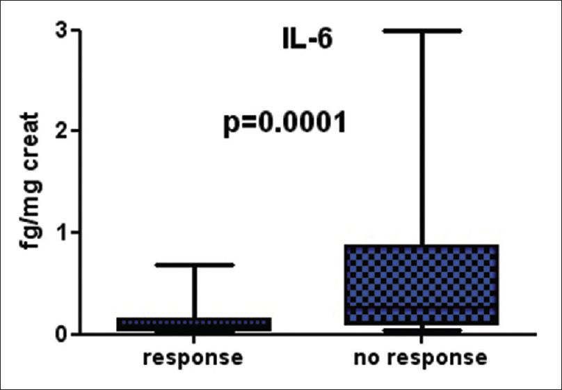
- Differences in interleukin-6 urinary levels in IgA nephropathy patients according to their response in treatment
Focal segmental necrotizing glomerulonephritis patients
Mean age of the patients with FSNGN (n = 40, males/females 24/16) at their presentation was 56.5 (range 25–80) years. Fourteen patients (35%) had PR3-ANCA, 22/40 (55%) had MPO-ANCA and 4/40 (10%) were ANCA negative. Mean levels of serum creatinine at presentation (Scr1) were 3.8 ± 2.5 mg/dl, CrCl1 23.68 ± 19.5 ml/min, and mean levels of proteinuria 1.5 ± 0.9 g/24 day. Eight patients presented with severe renal failure (CrCl1 < 10 ml/min/1.73 m2) requiring hemodialysis [Table 1].
Kidney biopsies showed global sclerosis in 20.4% ±19.8% of glomeruli, crescent formation in 68.9% ±25.9% of glomeruli, severe tubular atrophy (≥50% of tubules) in 16/40 (40%) patients. The only finding in renal histology which correlated significantly with renal function outcome was the percentage of crescents (r = −0.6, P = 0.003). Patients with crescents in ≥50% of glomeruli and patients with tubular atrophy in ≥50% of tubules had significantly increased urinary levels of MCP-1 (29 ± 32 vs. 0 fg/mg Ucr, P = 0.001 and 28.8 ± 10.7 vs. 0 fg/mg Ucr, P = 0.001, respectively) and TGF-β1 (0.9 ± 1.5 vs. 0.09 ± 0.09 fg/mg Ucr, P = 0.01 and 1.1 ± 1.5 vs. 0.15 ± 0.17 fg/mg Ucr, P = 0.002, respectively) [Figure 2]. There was no difference in urinary cytokine excretion between PR3 and MPO-ANCA (+) patients.
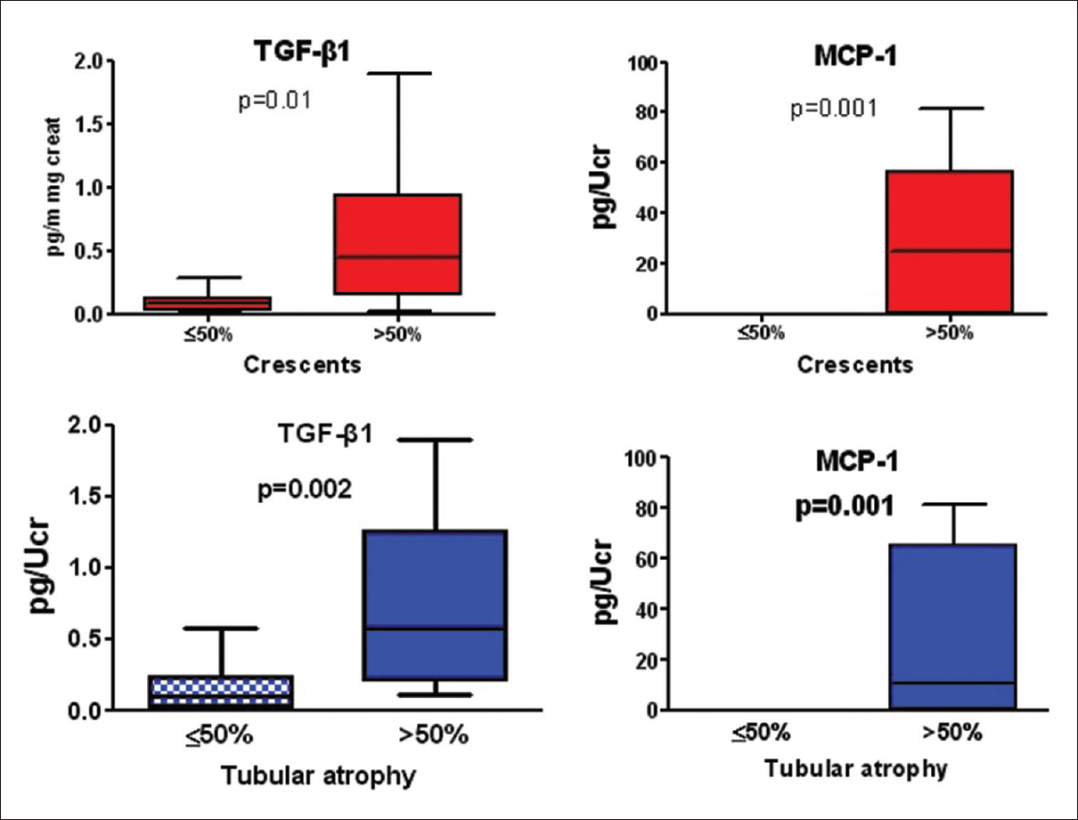
- Differences in urinary cytokine excretion in focal segmental necrotizing glomerulonephritis according to the severity of tubular atrophy and crescent formation
Compared to controls, FSNGN patients had significantly higher urinary levels of all cytokines and chemokines measured, while, in IgAN, only IL-2, IL-12, TNF-α, IL-6, IL-10, IL-17, MCP-1 and MIP-1 β urinary levels were higher (data not shown). Almost all cytokines and chemokines were significantly higher in FSNGN compared with IgAN patients as shown in Table 2.
Among FSNGN patients, MCP-1 urinary excretion had positive correlation with Th1 cytokines (IL-12, P = 0.009, TNF-α, P = 0.02, INF-γ, P = 0.03), Th2 cytokines (IL-4, P = 0.01, IL-6, P < 0.0001, IL-13, P = 0.03) and Treg/T17 cytokines (IL-17, P = 0.01, GM-CSF, P = 0.002) cytokines. The only independent parameter however for MCP-1 excretion was IL-17, r = 0.6, P = 0.04.
At the end of the follow-up period (44 months [1–180]), 24/40 patients (60%) responded to treatment, 4/40 (10%) had a declining renal function, not reaching ESRD and 12/40 patients (30%) developed ESRD and started on hemodialysis. Mean serum creatinine (Scr2) of the 28 patients, who remained off dialysis, was 1.7 ± 1 mg/dl and CrCl2 was 44.7 ± 23.9 ml/min/1.73 m2. Patients with FSNGN who responded to treatment had reduced Th2 (IL-4, IL-6), Treg (GM-CSF) and pro-inflammatory (MIP-1 β) chemokines [Figure 3].
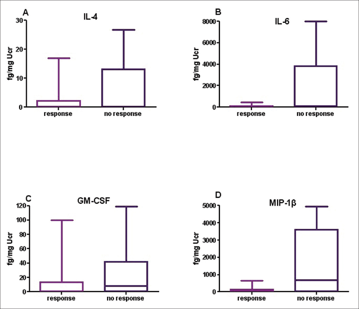
- Differences in Th1 and Th2 urinary cytokines in focal segmental necrotizing glomerulonephritis patients according to their response to treatment
Cytokine profile of patients who reached end-stage renal disease
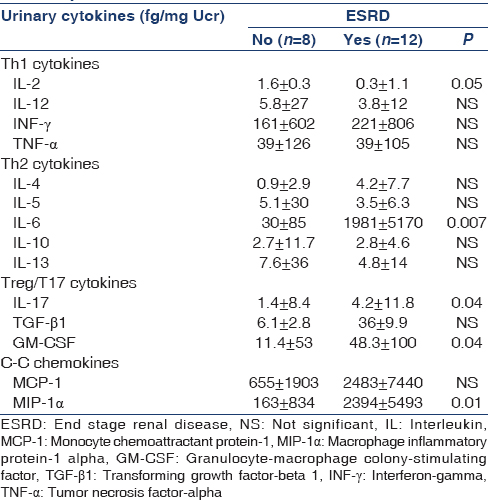
Urinary cytokine excretion was analyzed in terms of deterioration of renal function. Data in Table 5 demonstrate that patients, either diagnosed as IgAN or FSNGN, who finally reached ESRD had significantly increased excretion of IL-6, IL-17, GM-CSF and MIP-1 β, but reduced levels of IL-2.
Discussion
In the present study, we investigated the urinary cytokine excretion in two proliferative glomerulonephritides, IgAN, and FSNGN. Both diseases are characterized by proliferation of the native glomerular cells, together with macrophage and T cell infiltration. IgAN is characterized by immune complex deposition in the mesangium, followed by mesangial hypercellularity and occasionally endocapillary proliferation that progressively lead to glomerulosclerosis and tubular atrophy. Recent studies have demonstrated the presence of endocapillary proliferation as the most important histological finding associated with disease progression.[12131415] FSNGN is characterized by extracapillary proliferation and focal necrosis leading to end-stage renal disease if remained untreated.[16]
As expected, cytokine urinary excretion was significantly increased in FSNGN, compared to IgAN patients, and this difference was applied to almost all cytokines measured. Interestingly, even in patients with IgAN, those with crescent formation had significantly increased urinary excretion of IL-6, IL-10, MCP-1 and MIP-1 β, compared with IgAN patients without crescents in renal biopsy. The urinary cytokine excretion has been demonstrated to represent their local production within the kidneys in many different types of glomerulonephritis.[17] In IgAN, there was a strong correlation of both Th1 and Th2 cytokines with inflammatory infiltration and the degree of endocapillary proliferation. According to Oxford classification system, the presence of endocapillary proliferation is associated by more aggressive disease, as depicted by the degree of proteinuria, crescent formation and need for immunosuppressive treatment.[121318] Endocapillary proliferation in IgAN was shown to be accompanied by up-regulation of glomerular IL-10 mRNA expression.[19] In our study, the presence of endocapillary proliferation had significant correlation with the presence of extracapillary proliferation (crescents), and also with the urinary concentration of TNF-α, INF-γ, IL-6, IL-17, MCP-1 and MIP-1 β. To our knowledge, this is the first evidence that endocapillary proliferation was the main histologic finding correlated significantly with Th1, Th2, Th17 cytokines and also with chemokines. More interestingly, Th2 cytokines (IL-6, IL-10) and chemokines, but not Treg cytokines, had significant positive correlation with the presence of extracapillary proliferation, suggesting that interaction between IL-6 and chemokines predominates in the development and expansion of histological damage in IgAN. These results are in accordance with previous in vivo and in vitro studies. A recently described experimental model of IgAN in mice has developed after infection of mice with Mycoplasma penetrans, which, above the typical histology of IgAN, resulted in the increased expression of TNF-α, IL-6 and NF-kB.[20] In vitro studies have shown that stimulation of mesangial cells by galactose-deficient polymeric IgA1 results in enhanced secretion of IL-6.[21]
In contrast, in FSNGN the percentage of crescent formation had a positive correlation only with the preinflammatory chemokine MCP-1 and with Treg cytokine, TGF-β1, and no correlation was demonstrated between crescent formation and Th1 or Th2 cytokines. Both molecules also had a significant correlation with the degree of tubular atrophy. Involvement of C-C chemokines in glomerular macrophage accumulation and crescent formation in various forms of necrotising glomerulonephritis is well-established.[222324] In crescentic glomerulonephritis, MCP-1 is expressed by parietal epithelial cells, macrophages, infiltrating leucocytes, and also by tubular epithelial cells.[2225] MCP-1 is mainly chemoattractant for macrophages, and its blockade resulted in the reduction of macrophage infiltration. Previous studies have clearly demonstrated that in human crescentic glomerulonephritis, the majority of infiltrating cells within glomeruli are macrophages, which form the main cellular part of crescents,[2425] while T-cells are mainly located in the interstitium, not participating to the cellular composition of crescents.[21] However, the luck of a significant correlation between Th1 and Th2 urinary cytokines and severity of renal pathology does not imply a minor role of the above cytokines in disease progression. Their excess concentration in the urine and their significant correlation with the preinflammatory chemokines, MCP-1 and MIP-1 β, indicate that T-cell infiltration has a major contribution to renal pathology and may predispose to inflammatory process and macrophage accumulation.
However, the most important cytokine in our study was IL-17. IL-17 appeared to have a crucial and central role in the activation of the inflammatory process in both diseases, and also correlated with the development of ESRD. It was the only independent factor correlated with the presence of endocapillary proliferation in IgAN and with the MCP-1 urinary excretion in FSNGN. There are recent reports of IL-17 being involved in several forms of glomerulonephritis, including pauci-immune ANCA associated vasculitis, minimal change disease and IgAN.[102627] Cellular sources of IL-17 in ANCA associated glomerulonephritis are T-cells, mast cells and polymorphonuclear granulocytes which serve as IL-17 producers at the early stages of the disease.[26] IL-17 acts as a pleiotropic cytokine, chemoatractant for neutrophils and suppressor of Th1 and Treg cells.[2829] In the present study, the significant positive correlation between IL-17 and MCP-1 urinary excretion in FSNGN indicates that IL-17 may exert its pro-inflammatory effects by stimulating the production of MCP-1 and leading to macrophage accumulation.
Cytokines associated with the development of ESRD in patients, from both diseases, were IL-2, IL-6, IL-17, GM-CSF, and MIP-α. However, the way of intervention of cytokines had substantial differences in the two diseases. In FSNGN, increased urinary excretion of IL-4, IL-6, GM-CSF and MIP-1 β were associated with failure to response to immunosuppressive treatment, but Th1 cytokines had no significant involvement in disease outcome. In IgAN, a whole cohort of cytokines participated to renal impairment, although, in multiple regression analysis, IL-6 was the only independent factor correlated with CrCl at the end and with response to treatment. This is in agreement with previous studies, which showed that urinary excretion of IL-6 cytokine correlated with the severity of chronic histological lesions and outcome of renal function in IgAN.[6] The results of the present study suggest that IL-6 cytokine may have a global interactive role during the progression of renal damage in various forms of proliferative glomerulonephritis, and this role does not seem to be affected by current treatment strategies, such as prednisolone and immunosuppresives.
Conclusion
Our study demonstrated that urinary excretion of Th1, Th2 and Treg/Th17 cytokines were significantly higher in FSNGN compared to IgAN patients and had different roles in the two forms of glomerulonephritis. Both Th1 and Th2 cytokines were implicated in inflammatory reactions and renal pathology in IgAN while their contribution in histological changes in FSNGN appeared to be mediated through MCP-1 production. However, in both diseases, IL-17 was proved to be of particular interest in stimulating inflammation and IL-6 in leading to the progression of kidney injury.
Source of Support: Nil
Conflict of Interest: None declared.
References
- An update on the pathogenesis and treatment of IgA nephropathy. Kidney Int. 2012;81:833-43.
- [Google Scholar]
- Wegener's granulomatosis, microscopic polyarteritis and pauciimmune crescentic necrotizing glomerulonephritis, an overview. Przegl Lek. 1992;49:6-9.
- [Google Scholar]
- Morphologic markers of progressive immunoglobulin A nephropathy. Adv Chronic Kidney Dis. 2012;19:107-13.
- [Google Scholar]
- Detection of multiple cytokines in the urine of patients with focal necrotising glomerulonephritis may predict short and long term outcome of renal function. Cytokine. 2012;57:120-6.
- [Google Scholar]
- Urinary levels of epidermal growth factor, interleukin-6 and monocyte chemoattractant protein-1 may act as predictor markers of renal function outcome in immunoglobulin A nephropathy. Nephrology (Carlton). 2009;14:613-20.
- [Google Scholar]
- Chemokines and chemokine receptors in glomerulonephritis and renal allograft rejection. Med Sci Monit. 2007;13:RA31-6.
- [Google Scholar]
- Chemokines play a critical role in the cross-regulation of Th1 and Th17 immune responses in murine crescentic glomerulonephritis. Kidney Int. 2012;82:72-83.
- [Google Scholar]
- An essential role of interleukin-17 receptor signaling in the development of autoimmune glomerulonephritis. J Leukoc Biol. 2014;96:463-72.
- [Google Scholar]
- Imbalance of regulatory T cells to Th17 cells in IgA nephropathy. Scand J Clin Lab Invest. 2012;72:221-9.
- [Google Scholar]
- Review: T helper 17 cells: Their role in glomerulonephritis. Nephrology (Carlton). 2010;15:513-21.
- [Google Scholar]
- The Oxford IgA nephropathy clinicopathological classification is valid for children as well as adults. Kidney Int. 2010;77:921-7.
- [Google Scholar]
- The Oxford classification of IgA nephropathy: Pathology definitions, correlations, and reproducibility. Kidney Int. 2009;76:546-56.
- [Google Scholar]
- Validation of the Oxford classification of IgA nephropathy in cohorts with different presentations and treatments. Kidney Int. 2014;86:828-36.
- [Google Scholar]
- Validation of the Oxford classification of IgA nephropathy. Kidney Int. 2011;80:310-7.
- [Google Scholar]
- Clinicopathologic predictors of death and ESRD in patients with pauci-immune necrotizing glomerulonephritis. Am J Kidney Dis. 2003;41:29-37.
- [Google Scholar]
- MIP-1alpha and MCP-1 contribute to crescents and interstitial lesions in human crescentic glomerulonephritis. Kidney Int. 1999;56:995-1003.
- [Google Scholar]
- Clinicopathologic features and outcomes in endocapillary proliferative IgA nephropathy. Nephron Clin Pract. 2010;115:c161-7.
- [Google Scholar]
- In situ upregulation of IL-10 reflects the activity of human glomerulonephritides. Am J Kidney Dis. 1998;32:80-92.
- [Google Scholar]
- Mycoplasma penetrans infection is a potential cause of immunoglobulin A nephropathy: A new animal model. J Nephrol. 2013;26:470-5.
- [Google Scholar]
- The combined role of galactose-deficient IgA1 and streptococcal IgA-binding M Protein in inducing IL-6 and C3 secretion from human mesangial cells: Implications for IgA nephropathy. J Immunol. 2014;193:317-26.
- [Google Scholar]
- Expression of monocyte chemoattractant protein-1 in experimental crescentic glomerulonephritis in rats. J Lab Clin Med. 1997;129:239-44.
- [Google Scholar]
- Expression of the chemokine monocyte chemoattractant protein-1 and its receptor chemokine receptor 2 in human crescentic glomerulonephritis. J Am Soc Nephrol. 2000;11:2231-42.
- [Google Scholar]
- Glomerular expression of C-C chemokines in different types of human crescentic glomerulonephritis. Nephrol Dial Transplant. 2003;18:1526-34.
- [Google Scholar]
- Monoclonal antibody analysis of glomerular hypercellularity in human glomerulonephritis. Clin Nephrol. 1984;22:163-8.
- [Google Scholar]
- Renal IL-17 expression in human ANCA-associated glomerulonephritis. Am J Physiol Renal Physiol. 2012;302:F1663-73.
- [Google Scholar]
- Increased urinary excretion of interleukin-17 in nephrotic patients. Nephron. 2002;91:243-9.
- [Google Scholar]
- TGF-beta-induced Foxp3 inhibits T (H) 17 cell differentiation by antagonizing RORgammat function. Nature. 2008;453:236-40.
- [Google Scholar]
- Myelin-specific regulatory T cells accumulate in the CNS but fail to control autoimmune inflammation. Nat Med. 2007;13:423-31.
- [Google Scholar]


